Download

1 / 72
740 likes | 854 Vues
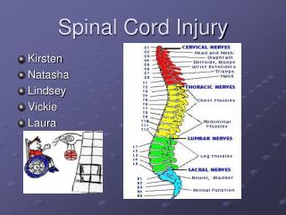
Respiratory disease main cause of death in Spinal Cord Injury. A & P Refresher Acute phase Respiratory Physio Techniques Weaning Cardiovascular Tracheostomies Prognosis. 68 patients >C5 88% needed intubating C5-C8 60% needed intubating. Velmahos gc et al American surgeon 2003.

E N D
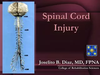
Respiratory disease main cause of death in Spinal Cord Injury
A & P Refresher • Acute phase • Respiratory • Physio Techniques • Weaning • Cardiovascular • Tracheostomies • Prognosis
68 patients >C5 88% needed intubating C5-C8 60% needed intubating Velmahos gc et al American surgeon 2003 156 Patients Injuries C2-C8 107 required tracheostomies Harop et al Journal of neurosurgery spine 2004 Respiratory compromise Level of injury Age Premorbid resp. disease
MAG (myelin-associated glycoprotein), Omgp (oligodendrocyte myelin glycoprotein), KDI (synthetic: Lysine–Asparagine–Isoleucine ‘g-1 of Laminin Kainat Domain’), Nogo (Neurite outgrowth inhibitor), NgR (Nogo protein Receptor), the Rho signaling pathway (superfamily of ‘Rho-dopsin gene including neurotransmitter receptors‘), EphA4 (Ephrine), GFAP (Glial Fibrillary Acidic Protein), different subtypes of serotonergic and glutamatergic receptors, antigens, antibodies, immune modulators, adhesion molecules, scavengers, neurotrophic factors, enzymes, hormones, collagen scar inhibitors, remyelinating agents and neurogenetic/plasticity inducers Pathophysiology Trauma ↓ Haemorrhage/Inflammatory mediators ↓ Oedema ↓ Ischaemia ↓ Oedema ↓ Ischaemia ↓ Oedema ↓ Ischaemia ↓
Cardiorespiratory physiology
Respiratory Afferents Intrapulmonary receptors Vagus Stretch/proprioreceptors ribs/intercostals T1-T12 Clavicles Low Cervical Chemoreceptors Carotid body Chemoreceptors Brainstem
Acute changes Damaged cord becomes unresponsive Flaccid, areflexic Lasts for 6 days to 6 weeks
Respiratory • Can’t breath • Can’t cough
Acute VC 1 Year VC Lumbar Unable to cough 100-70% 100-70% Low thoracic é chest wall compliance ê Vital capacity High thoracic éé chest wall compliance 30-50% êê Vital capacity poor expansion. Basal collapse 60-70% C5/C6 Diaphragms, Scalenes, 20% 40-50% C3/C4/C5 Sternomastoid and partial diaphragm Above C3 Sternomastoid only 5-10%
Acute changes respiratory autonomic Bronchial hypersecretion Bronchial hyper-responsiveness
Not forgetting… Head injuries Chest wall trauma Pulmonary contusion Haemopneumothorax PE / Fat embolus
Acute Respiratory monitoring Lung function FVC, PEFR, Speech, RR, Resp Pattern FVC> 1L FVC < 1L FVC= Tidal volume Pulse oximeter Blood gases Watch closely in an appropriate environment for several days
Acute Respiratory Treatment Oxygen A good physiotherapist !
Early Respiratory System Complications Atelectasis Hypersecretion Bronchospasm Pulmonary Oedema Pneumonia Chest Trauma Respiratory Failure Pulmonary Thromboembolism
Respiratory assessment • FVC • Observations - mode of ventilation, FiO2, SaO2, RR • ABGs, CVS • CXR • Auscultation • Cough?
Observation of breathing pattern Paradoxical breathing Unilateral breathing Abdominal breathing Respiratory rate Cough
Importance of FVC • Around or less than 1L
Non Invasive Management? • Regular FVC • Chest physiotherapy • Cough assist + manual techniques • IPPB with the nurses • Spinal stability? • Nutrition? • Don’t wait to intubate if it is inevitable…
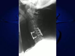
Intubation? • The Neurological level of Injury and completeness of injury are the most important predictors of requirement for tracheostomy • Early semi-elective intubation during the day by senior experienced staff is preferable to emergency intubation • Care should be taken when considering extubation of high cervical cord injured patients following stabilisation surgery
Ventilation? • Some evidence that higher inspiratory pressures reduce the effects of atelectasis • Rather than a high PEEP • PEEP aim for 5 cmH2O • ETv around 500ml or 15-20ml/kg • NICE Guideline 6-8ml/kg LPV
Secretion management • Carbocysteine • N acetylcysteine nebs • Saline nebs ? • Bronchodilator nebs • Hyoscine? • Azithromycin / colistin nebs for colonisation • Supraglottic suction tubes
Positioning: Supine vs Sitting • FVC must test in supine • In head tilt down increases by 6% • Sat upright decreases by 14% • Use of a binder helps in sitting • Roll your patients… • Combine therapy with nursing requirements
Aggressive Management of Atelectasis • Expansion / loosening of secretions to reduce mucus plugging • Use of ‘sighs’ within Mechanical Ventilation • Four hourly bronchodilation, heated humidification & Mucolytics • The Vest? • Intrapulmonary Percussive Ventilation?
Respiratory techniques • Suctioning - unopposed vagal stimulation: atropine nearby • Expiratory vibs / shakes / percussion • The Cough Assist Machine? • Assisted cough • MHI • Inspiratory Muscle Training • VFB/Weaning
Please Do… • ASIA charting • Refer to MASCIP guidelines for moving & handling • Positioning and skin care • Pressure care mattress • Bowel routine: More MASCIP guidelines • Limb care
Please Don’t… • Sit patients up - yet • Use a Tilt Table – yet • Sit your patient on the edge of the bed – ever!
Ventilated spinal injured patients • 15-20% Initially ventilated • 98% Weanable • 1% Nocturnal ventilation • 1% Fully ventilator dependant • = 8-12 patients/yr • ~ 120 patients in UK
Weaning Based on little evidence but vast experience Prerequisites Good pulmonary compliance Low FiO2 requirement Awake and cooperative Some respiratory activity Committed team
Any respiratory activity? Testing Volume measurement Beware sensitive ITU Vents Modified brainstem death test
Weaning Progressive ventilator free breathing Measure Vital Capacity VC Time off Vent <250 mls 5 Mins -500 mls 15 Mins -750 mls 30 Mins -1000 mls 60 Mins Measure VC Post weaning >70% pre weaning Southport Spinal Injury Centre Increase duration and/ or frequency
Weaning Wait for spasticity Bronchodilators ?High TV Ventilation (>20 ml/Kg)?1 Supine • The effect of tidal volumes on the time to wean persons with high tetraplegia from ventilators • Peterson W. et al spinal cord 1999 37(4):284-288
Weaning Off vent requires PEEP/CPAP to reduce atalectasis Best option cuff with speaking valve. Ditch the ITU vent Don’t reduce pressure support too far Try to stick to plan Aim for off all day, support at night
Speech essential Eating optional
How to wean BIPAP/ PS Slow weaners Fast weaners laryngeal function vs resp function VFB Cuff up Cuff down on vent VFB Cuff down speaking valve VFB speaking valve Downsized uncuffed tube Decannulate
How successful ? Southport spinal injuries unit • 246 patients over 20 years • 63% weaned • 33% Ventilator dependant • 4% Died
Post weaning Maintenance ‘ Maintain Range of Movements’ Manual hyperinflation IPPB Cough Assist/ Clearway Improve muscle strength Inspiratory muscle training
Cardiovascular • Can’t squeeze • Can’t speed up
Parasympathetic Sympathetic Parasympathetic Vasoconstriction Vasodilation T6 Balance point Hypotension, bradycardia, tendency to asystole