Download

1 / 41
420 likes | 580 Vues
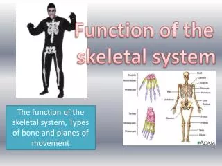
The skeletal system Structure and function of bone Organization of the skeleton Joints. Functions of bone (skeleton) Support and protection Blood cell formation Mineral storage (calcium especially) Site for muscle attachment body movement Protect blood vessels, nerves.

E N D
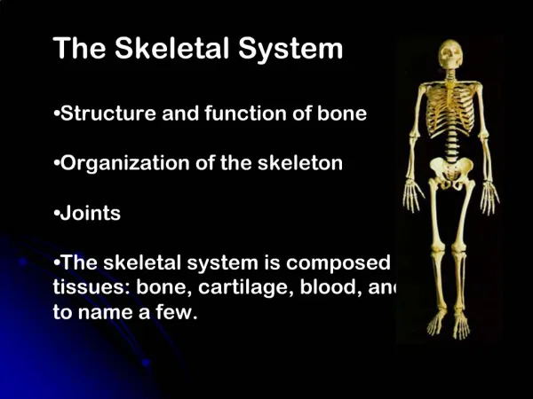
The skeletal system Structure and function of bone Organization of the skeleton Joints
Functions of bone (skeleton) Support and protection Blood cell formation Mineral storage (calcium especially) Site for muscle attachmentbody movement Protect blood vessels, nerves
Bones classified by shape: long, short, flat, irregular, round Long bones- in limbs Short bones- wrists and ankles Flat bones- ribs, scapulae, cranium Irregular- vertebrae, facial bones Sesamoid (round)- patella
Compact bone osteocytes within lacunae arranged in concentric circles called lamellae This surrounds a central canal; complex is called osteon (Haversian system) Canaliculi connect osteocytes to central canal and to each other
Prenatal development skeleton is mostly cartilaginous Cartilage cells and then osteoblasts start to deposit minerals Cartilaginous disk (epiphyseal disk) remains in epiphysis Cells eventually stop dividing
Fetal bone development • Intramembranous-sheets of connective tissue • Endochondrial- from masses of hyaline cartilage
Adults continually break down (resorb) and build up (deposit) bone Osteoclasts remove damaged cells and release calcium into blood osteoclasts come from bone marrow Osteoblasts remove calcium from blood and build new matrix. They become trapped osteocytes osteoblasts originate in connective tissue
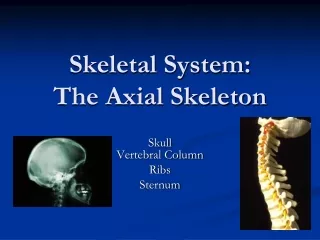
Axial skeleton skull (cranium and facial bones) hyoid bone (anchors tongue and muscles associated with swallowing) vertebral column (vertebrae and disks) thoracic cage (ribs and sternum) Appendicular skeleton pectoral girdle (clavicles and scapulae) upper limbs (arms) pelvic girdle (coxal bones, sacrum, coccyx) lower limbs (legs)
posterior view p. 135
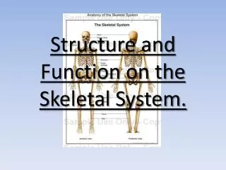
Bones named and numbered in Table 7.1 Terms listed in table 7.2 Axial skeleton supports and protects organs of head, neck and trunk Appendicular skeleton- bones of limbs and bones that anchor them to the axial skeleton Articulation- where joints are formed
22 bones in skull 6 in middle ears 1 hyoid bone 26 in vertebral column 25 in thoracic cage 4 in pectoral girdle 60 in upper limbs 60 in lower limbs 2 in pelvic girdle 206 bones in all
The skull 8 sutured bones in cranium Facial bones: 13 sutured bones, 1 mandible Cranium encases brain attachments for muscles sinuses
Allows for growth
Vertebral column 7 cervial vertebrae 12 thoracic 5 lumbar 1 sacrum (5 fused 1 coccyx (4 fused) Vertebrae vary in size and morphology
Thoracic cage ribs thoracic vertebrae sternum costal cartilages True ribs are directly attached to the sternum (first seven pairs) Three false ribs are joined to the 7th rib Two pairs of floating ribs
Clavicles and scapulae Help brace shoulders Attachment sites for muscles
Bones of upper limb Humerus (upper arm) Radius; ulna Carpals, metacarpals, phalanges Bones of lower limb Femur Patella Tibia, fibula Tarsals, metatarslas, phalanges
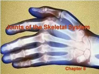
Joints Immovable (synarthoses) bones sutured together by connective tissue: skull Slightly movable (amphiarthoses) connected by fibrocartilage or hyaline cartilage: vertebrae, rib/sternum joint, pubic symphysis Freely movable (diarthroses)- separated ligaments- hold bones together tendons- muscle to bone lined by synovial membrane
Different types of synovial joints Condylar Hinge Saddle Ball-and-socket Plane Pivot
Types of movement and examples (with muscles) flexion- move lower leg toward upper extension- straightening the leg abduction- moving leg away from body adduction- movong leg toward the body rotation- around its axis supination- rotation of arm to palm-up position pronation- palm down circumduction- swinging arms in circles inversion- turning foot so sole is inward eversion- sole is out
Elevation and depression- raising body part up or down Aging and bones both bone and cartilage tend to deteriorate cartilage: chondrocytes die, cartilage becomes calcified osteoporosis; bone is broken down faster than it can be built bones get weak and brittle; tend to fracture easily
Risk factors for osteoporosis Inadequate calcium Little weight-bearing exercise Drinking alcohol, smoking Being female: decreased estrogen secretion after menopause Small frame Caucasian or Asian ethnicity
Skeleton and other systems Skin makes vitamin D which enhances calcium absorption Skeleton stores calcium for muscle contraction, nervous stimulation, blood clot formation Red marrow- site of blood cell formation Calcium levels regulated by parathyroid hormone and calcitonin kidneys (can help provide vitamin D) digestive system (can release calcium into blood
Regulation of blood calcium levels
Growth hormone regulates skeletal growth stimulates cell division in epiphyseal disks in long bones Growth stops when epiphyseal disks are converted to bone When excess growth hormone is produced in childhoodgigantism In adulthood- acromegaly. Bones can’t grow but soft tissue can
This man suffered from a pituitary tumor
When muscle contracts, it shortens and causes movement Skeletal muscles attached to bones by tendons Insertion- attachment to more movable bone Origin- less movable Flexors and extensors act on the same joint to produce opposite actions You need both bones and muscles for movement!