Chapter 7 Bone Tissue
Chapter 7 Bone Tissue. Tissues and organs of the skeletal system Histology of osseous tissue Bone development Physiology of osseous tissue Bone disorders. Bone as a Tissue. Dynamic tissue that continually remodels itself Bones and bone tissue

Chapter 7 Bone Tissue
E N D
Presentation Transcript
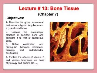
Chapter 7Bone Tissue • Tissues and organs of the skeletal system • Histology of osseous tissue • Bone development • Physiology of osseous tissue • Bone disorders
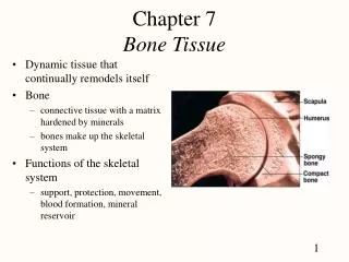
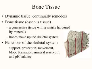
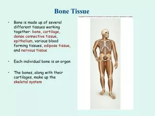
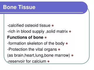
Bone as a Tissue • Dynamic tissue that continually remodels itself • Bones and bone tissue • bone or osseous tissue is a connective tissue with a matrix hardened by minerals(calcium phosphate) • bones make up the skeletal system • individual bones are made up of bonetissue, marrow, cartilage & periosteum • Functions of the skeletal system • support, protection, movement, blood formation, mineral reservoir, pH balance & detoxification
Structure of a Flat Bone • External and internal surfaces of flat bone are composed of compact bone • Middle layer is spongy bone (diploe). No marrow cavity • Blow to the skull may fracture outer layer and crush diploe, but not harm inner compact bone
Structure of a Long Bone • Periosteum & articular cartilage • Compact & spongy bone • Endosteum • Yellow marrow
General Features of Bones • Shaft (diaphysis) is cylinder of compact bone containing marrow cavity (medullary cavity) & lined with endosteum (layer of osteopenic cells and reticular connective tissue) • Enlarged ends (epiphyses) are spongy bone covered with a layer of compact bone • enlarged to strengthen joint & provide for attachment of tendons and ligaments • Joint surface covered with articular cartilage (lubrication) • Remainder of bone covered with periosteum • outer fibrous layer of collagen fibers continuous with tendons or perforating(Sharpey’s) fibers that penetrate into bone matrix • inner osteogenic layer important for growth & healing • Epiphyseal plate or line depends on age
Cells of Osseous Tissue (1) • Osteogenic cells reside in endosteum, periosteum or central canals • arise from embryonic fibroblasts and become only source for new osteoblasts • multiply continuously & differentiate into amitotic osteoblasts in response to stress or fractures • Osteoblasts form and help mineralize organic matter of matrix • Osteocytes are osteoblasts that have become trapped in the matrix they formed • cells in lacunae connected by gap junctions inside canaliculi • signal osteoclasts & osteoblasts about mechanical stresses
Cells of Osseous Tissue (2) • Osteoclasts develop in bone marrow by the fusion of 3-50 of the same stem cells that give rise to monocytes found in blood • Reside in pits called resorption bays that they have eaten into the surface of the bone
Matrix of Osseous Tissue • Dry weight is 1/3 organic & 2/3 inorganic matter • Organic matter • collagen, glycosaminoglycans, proteoglycans & glycoproteins • Inorganic matter • 85% hydroxyapatite (crystallized calcium phosphate salt) • 10% calcium carbonate • other minerals (fluoride, sulfate, potassium, magnesium) • Combination provides for strength & resilience • minerals resist compression; collagen resists tension • fiberglass = glass fibers embedded in a polymer • bone adapts to tension and compression by varying proportions of minerals and collagen fibers
Compact Bone • Osteon (haversian system) = basic structural unit • cylinders of tissue formed from layers (lamellae) of matrix arranged around central canal holding a blood vessel • collagen fibers alternate between right- and left-handed helices from lamella to lamella • osteocytes connected to each other and their blood supply by tiny cell processes in canaliculi • Perforating canals or Volkmann canals • vascular canals perpendicularly joining central canals • Circumferential or outer lamellae
Spongy Bone • Spongelike appearance formed by rods and plates of bone called trabeculae • spaces filled with red bone marrow • Trabeculae have few osteons or central canals • no osteocyte is far from blood of bone marrow • Provides strength with little weight • trabeculae develop along bone’s lines of stress
Bone Marrow • Soft tissue that occupies the medullary cavity of a long bone or the spaces amid the trabeculae of spongy bone • Red marrow looks like thick blood • mesh of reticular fibers and immature cells • hemopoietic means produces blood cells • found in vertebrae, ribs, sternum, pelvic girdle and proximal heads of femur and humerus in adults • Yellow marrow • fatty marrow of long bones in adults • Gelatinous marrow of old age • yellow marrow replaced with reddish jelly
Intramembranous Ossification • Produces flat bones of skull & clavicle • Steps of the process • mesenchyme condenses into a sheet of soft tissue • transforms into a network of soft trabeculae • osteoblasts gather on the trabeculae to form osteoid tissue (uncalcified bone) • calcium phosphate is deposited in the matrix transforming the osteoblasts into osteocytes • osteoclasts remodel the center to contain marrow spaces & osteoblasts remodel the surface to form compact bone • mesenchyme at the surface gives rise to periosteum
Endochondral Ossification • Primary ossification center forms in cartilage model • chondrocytes near the center swell to form primary ossification center • matrix is reduced & model becomes weak at that point • cells of the perichondrium produce a bony collar • cuts off diffusion of nutrients and hastens their death • Primary marrow space formed by periosteal bud • osteogenic cells invade & transform into osteoblasts • osteoid tissue deposited and calcified into trabeculae at same time osteoclasts work to enlarge the primary marrow cavity
Primary Ossification Center & Marrow Space • Both form in center of cartilage model -- same process begins again subsequently at ends of cartilage model.
The Metaphysis • Transitional zone between head and shaft of a developing long bone • Zone of reserve cartilage is layer of resting cartilage • Zone of cell proliferation is layer • chondrocytes multiply forming columns of flat lacunae • Zone of cell hypertrophy shows hypertrophy • Zone of calcification shows mineralization between columns of lacunae • Zone of bone deposition -- chondrocytes die and each channel is filled with osteoblasts and blood vessels to form a haversian canal & osteon
Secondary Ossification Center • Begin to form in the epiphyses near time of birth • Same stages occur as in primary ossification center • result is center of epiphyseal cartilage being transformed into spongy bone • Hyaline cartilage remains on joint surface as articular cartilage and at junction of diaphysis & epiphysis (epiphyseal plate) • each side of epiphyseal plate has a metaphysis
Metaphysis & Secondary Ossification Center • Metaphysis is cartilagenous material that remains as growth plate between medullary cavity & secondary ossification centers in the epiphyses.
Bone Growth and Remodeling • Grow and remodel themselves throughout life • growing brain or starting to walk • athletes or history of manual labor have greater density & mass of bone • Cartilage grows by both appositional & interstitial growth • Bones increase in length by interstitial growth of epiphyseal plate • Bones increase in width by appositional growth • osteoblasts lay down matrix in layers parallel to the outer surface & osteoclasts dissolve bone on inner surface • if one process outpaces the other, bone deformities occur(osteitis deformans)
Achondroplastic Dwarfism • Short stature but normal-sized head and trunk • long bones of the limbs stop growing in childhood but other bones unaffected • Result of spontaneous mutation when DNA is replicated • mutant allele is dominant • Pituitary dwarf has lack of growth hormone • short stature with normal proportions
Mineral Deposition • Mineralization is crystallization process in which ions (calcium, phosphate & others) are removed from blood plasma & deposited in bone tissue • Steps of the mineralization process • osteoblasts produce collagen fibers that spiral along the length of the osteon in alternating directions • fibers become encrusted with minerals hardening matrix • ion concentration must reach the solubility product for crystal formation to occur & then positive feedback forms more • Ectopic ossification is abnormal calcification • may occur in lungs, brain, eyes, muscles, tendons or arteries (arteriosclerosis)
Mineral Resorption • Process of dissolving bone & releasing minerals into the blood • performed by osteoclasts “ruffled border” • hydrogen pumps in the cell membrane secrete hydrogen ions into the space between the osteoclast & the bone • chloride ions follow by electrical attraction • hydrochloric acid with a pH of 4 dissolves bone minerals • an enzyme (acid phosphatase) digests the collagen • Dental braces reposition teeth, creating greater pressure on the bone on one side of the tooth and less on the other side • increased pressure stimulates osteoclasts; decreased pressure stimulates osteoblasts to remodel jaw bone
Functions of Calcium & Phosphate • Phosphate is a component of DNA, RNA, ATP, phospholipids, & acid-base buffers • Calcium is needed for communication between neurons, muscle contraction, blood clotting & exocytosis • Calcium plasma concentration is 9.2 to 10.4 mg/dL -- 45% is as Ca+2, rest is bound to plasma proteins & is not physiologically active • Phosphate plasma concentration is 3.5 to 4.0 mg/dL & occurs in 2 forms: HPO4-2 & H2PO4-
Ion Imbalances • Changes in phosphate concentration have little effect • Changes in calcium can be serious • hypocalcemia is deficiency of blood calcium • causes excessive excitability of nervous system leading tomuscle spasms, tremors or tetany • laryngospasm may cause suffocation • calcium normally binds to cell surface contributing to resting membrane potential • with less calcium, sodium channels open more easily exciting neuron • hypercalcemia • excessive calcium binding to cell surface makes sodium channels less likely to open, depressing nervous system • Calcium phosphate homeostasis depends on calcitriol, calcitonin & PTH
Carpopedal Spasm • Hypocalcemia causing overexcitability of nervous system and muscle spasm of hands and feet
Calcitriol (Activated Vitamin D) • Produced by the following process • UV radiation penetrating the epidermal keratinocytes converts a cholesterol derivative (7-dehydrocholesterol) to previtamin D3 and then (cholecalciferol) D3 • liver adds OH to convert it to calcidiol • kidney adds OH to convert calcidiol to calcitriol • Calcitriol behaves as a hormone (blood-borne messenger) • stimulates intestine to absorb calcium, phosphate & magnesium • weakly promotes urinary reabsorption of calcium ions • promotes osteoclast activity to raise blood calcium concentration to the level needed for bone deposition • Abnormal softness of the bones is called rickets in children and osteomalacia in adults
Hormonal Control of Calcium Balance • Calcitriol, PTH and calcitonin maintain normal blood calcium concentration.
Calcitonin • Secreted by C cells of the thyroid gland when calcium concentration rises too high • Functions • reduces osteoclast activity by as much as 70% in 15 minutes • increases the number & activity of osteoblasts • Important role in children, but little effect in adults • calcitonin deficiency is not known to cause any disease in adults • may be useful in reducing bone loss in osteoporosis
Parathyroid Hormone • Secreted by the parathyroid glands found on the posterior surface of the thyroid gland • Released when calcium blood level is too low • Functions • binds to osteoblasts causing them to release osteoclast-stimulating factor that stimulates osteoclast multiplication & activity • promotes calcium resorption by the kidneys • promotes calcitriol synthesis in the kidneys • inhibits collagen synthesis and bone deposition by osteoblasts • Injection of low levels of PTH can cause bone deposition
Other Factors Affecting Bone • 20 or more hormones, vitamins & growth factors not well understood • Bone growth especially rapid at puberty • hormones stimulate proliferation of osteogenic cells and chondrocytes in growth plate • adolescent girls grow faster than boys & reach their full height earlier (estrogen has stronger effect) • males grow for a longer time • Growth ceases when epiphyseal plate “closes” • anabolic steroids may cause premature closure of growth plate producing short adult stature
Fractures and Their Repair • Stress fracture is a break caused by abnormal trauma to a bone • car accident, fall, athletics, etc • Pathological fracture is a break in a bone weakened by some other disease • bone cancer or osteoporosis • Fractures are classified by their structural characteristics -- causing a break in the skin, breaking into multiple pieces, etc • or after a physician who first described it
Healing of Fractures • Normally healing takes 8 - 12 weeks (longer in elderly) • Stages of healing • fracture hematoma (1) • broken vessels form a blood clot • granulation tissue (2) • fibrous tissue formed by fibroblasts & infiltrated by capillaries • callus formation (3) • soft callus of fibrocartilage replaced by hard callus of bone in 6 weeks • remodeling (4) occurs over next 6 months as spongy bone is replaced with compact bone
Healing of Fractures 1 2 3 4
Treatment of Fractures • Closed reduction • fragments are aligned with manipulation & casted • Open reduction • surgical exposure & repair with plates & screws • Traction is not used in elderly due to risks of long-term confinement to bed • hip fractures are pinned & early walking is encouraged • Electrical stimulation is used on fractures that take longer than 2 months to heal • Orthopedics = prevention & correction of injuries and disorders of the bones, joints & muscles
Osteoporosis • Most common bone disease • Bones lose mass & become brittle due to loss of both organic matrix & minerals • risk of fracture of hip, wrist & vertebral column • lead to fatal complications such as pneumonia • widow’s (dowager’s) hump is deformed spine • Postmenopausal white women at greatest risk • by age 70, average loss is 30% of bone mass • ERT slows bone resorption, but best treatment is prevention -- exercise & calcium intake (1000 mg/day) between ages 25 and 40 • Therapies to stimulate bone deposition are still under investigation