Efficient Loading of SPIO Nanoparticles in MSC and BMMC Visualized by Multi-Channel Flow Cytometry
This study demonstrates the effective incorporation of SPIO nanoparticles and CMTMR fluorescence into Mesenchymal Stem Cells (MSC) and Bone Marrow Mononuclear Cells (BMMC). Utilizing phase contrast microscopy, CMTMR, and Dragon Green fluorescence, micrographs reveal significant internalization of these particles. Flow cytometry analysis confirms that over 90% of the cells were successfully co-labeled with Dragon Green-SPIO and CMTMR, showcasing the proficiency of this loading technique for potential applications in cellular imaging and therapeutic interventions.

Efficient Loading of SPIO Nanoparticles in MSC and BMMC Visualized by Multi-Channel Flow Cytometry
E N D
Presentation Transcript
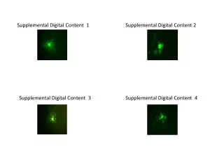
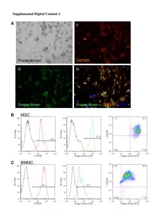
Supplemental Digital Content 2 A i ii Phase contrast CMTMR iii iv Dragon Green Dragon Green +CMTMR+DAPI B MSC C BMMC
Figure 2. Efficiency of SPIO nanoparticle loading into MSC and BMMC. (A) Micrographs of MSC showing incorporated SPIO by (i) phase contrast, (ii) CMTMR fluorescence, (iii) Dragon-Green fluorescence and (iv) a merged image of Dragon Green, CMTMR and DAPI nuclear label. Flow cytometric analysis of (B) MSC and (C) BMMC labeled with CMTMR (left panels) and Dragon Green-SPIO (centre panels). Proficient loading of MSC and BMMC with both SPIO nanoparticles and CMTMR was confirmed by multi-channel flow cytometry (right panels), which revealed >90% Dragon Green-SPIO and CMTMR colabeled cells.