Peripheral Vascular System: Anatomy & Health Guidelines
500 likes | 829 Vues
Learn about arteries, veins, and lymphatic system in the peripheral vascular system. Discover symptoms, diseases, and techniques for examination and health promotion.

Peripheral Vascular System: Anatomy & Health Guidelines
E N D
Presentation Transcript
Anatomy and Physiology - Arteries • Arterial pulses are palpable when artery lies close to body surface • Arms – Two/Three Locations • Brachial: above bend elbow • Radial: flexor surface wrist laterally • Ulnar: overlying tissues frequently obscure • Legs – Four Locations • Femoral: below inguinal ligament • Popliteal: behind knee • Dorsalis pedis: dorsum foot; lateral extensor tendon • Posterior tibial: behind medial malleolus ankle
Anatomy and Physiology - Veins • Deep Veins • Carry ~ 90% venous return from lower extremities • Superficial Veins: subcutaneous • Communicating Veins • Connect superficial (saphenous) system with deep system • Deep, superficial and communicating veins have one-way valves • Allow venous blood to flow from superficial to deep system toward heart, but not in opposite direction
Anatomy and Physiology – Lymphatic System • Extensive vascular network that drains lymph from bodily tissue and returns it to venous circulation • Lymph nodes • Round, oval or bean-shaped structures • Vary in size according to location • Important role in body’s immune system • Cells in lymph nodes engulf cellular debris/bacteria and produce antibodies • Only superficial lymph nodes accessible to physical examination
Health History • Common or Concerning Symptoms • Pain in arms/legs • Intermittent claudication • Cold, numbness, pallor in legs, hair loss • Color change in fingertips or toes in cold weather • Swelling in calves, legs or feet • Swelling with redness or tenderness
Health History • Arterial Peripheral Vascular Disease Legs • Intermittent claudication • Ask patients “Have you ever had any pain/cramping in legs when walking or with exercise?”; “Does pain get better with rest?” • Arterial spasm Fingers/Toes • Ask patients “Do your fingertips/toes ever change color in cold weather or when you handle cold objects?” • Venous peripheral vascular disease • Swelling of feet and legs • Ask about ulcers on lower legs, often near the ankles
Health Promotion and Counseling • Most patients with peripheral arterial disease (PAD) have no symptoms or non-specific symptoms • Triad of exercise-induced calf pain that causes stopping of exercise and relief of pain in 10 minutes present in only 10% affected patients • Screen for subclinical PAD • Aggressive risk factor intervention
Health Promotion and Counseling • Risk factors for Peripheral Arterial Disease • Current/past tobacco use • Hyperlipidemia • Diabetes mellitus • CAD • Hypertension • Cerebrovascular disease
Ankle-Brachial Index (ABI) • Detects stenosis of 50% or more in major vessels of legs • Measure systolic blood pressure (with Doppler ultrasonography) in each arm and in dorsalispedis and posterior tibial pulses • Calculate reading for right and left • Divide arm pressure by ankle pressure • ABI 0.90-1.30: normal ABI 0.41-.90: mild to moderate disease ABI 0.00-0.40: severe disease with critical stenosis
Techniques of Examination • Important Areas of Examination • Arms • Size, symmetry, skin color • Radial pulse, brachial pulse • Epitrochlear lymph nodes • Legs • Size, symmetry, skin color • Femoral pulse and inguinal lymph nodes • Popliteal, dorsalis pedis and posterior tibial pulses • Peripheral edema
Techniques of Examination - Arms • Inspect both arms from fingertips to shoulders • Note: • Size, symmetry and any swelling • Venous pattern • Color of skin and nail beds; texture of skin • Palpate radial pulse • Use pads of fingers on flexor surface of wrist • Partially flex patient’s wrist • Compare pulse in both arms
Techniques of Examination - Arms • Palpate Brachial Pulse • Flex elbow slightly • Palpate artery medial to biceps tendon in antecubital crease • Epitrochlear nodes • Flex elbow 90° • Support forearm • Feel in groove between biceps and triceps muscle, 3 cm above medial epicondyle
Techniques of Examination - Legs • Patient should lay down, draped so external genitalia covered and legs fully exposed • MUST remove stockings or socks • Inspect both legs from groin and buttocks to feet • Note: • Size, symmetry and any swelling • Venous pattern/venous enlargement • Pigmentation, rashes, scars or ulcers • Color and texture of skin, color of nail beds, distribution of hair on lower legs, feet and toes
Techniques of Examination - Legs • Palpate superficial inguinal nodes • Horizontal/Vertical groups • Note size, consistency and discreteness and tenderness • Nontender, discrete nodes up to 1-2cm are palpable in normal people
Palpate for the superficial inguinal nodes • Horizontal group: below the inguinal legement • Vertical group: near the upper part of the saphenous vein
Techniques of Examination – Palpate Pulses • Femoral Pulse • Press deeply below inguinal ligament • Midway between anterior superior iliac spine and symphysis pubis • Popliteal Pulse • Flex knee some, leg relaxed • Place fingertips of both hands to meet midline behind knee and press deeply into popliteal fossa • Dorsalis Pedis Pulse • Feel dorsum of foot, lateral to extensor tendon of great toe • Posterior tibial pulse • Curve fingers behind, slightly below medial malleolus of ankle
Palpate the popliteal pulse • In the tissue behind the knee
palpate the dorsalis pedis pulse • Dorsum of the foot, just lateral to the extensor tendon of the big toe.
Palpate the posterior tibial pulse • Behind the medial malleoulus of the ankle.
Grading Amplitude of Arterial Pulses • 4+ Bounding • 3+ Increased • 2+ Brisk, expected • 1+ Diminished, weaker than expected • 0 Absent, unable to palpate
Techniques of Examination - Edema • Compare one foot and leg with the other • Note relative size and prominence of veins, tendons and bones • Check for pitting edema • Press firmly with thumb for 5 seconds over dorsum each foot, behind medial malleolus and shins • Look for pitting (depression caused by pressure from thumb) • Severity of edema graded on four-point scale (slight to very marked)
Grade edema • May be graded by measuring the depth of pitting in centimeters, or by weight change, or the time pitting remains after releasing the pressure. • Grade edema on this scale • 1+: Mild pitting, slight indentation, no perceptible swelling of the leg • 2+: Moderate pitting, indentation subsides rapidly • 3+: Deep pitting, indentation remains for a short time, leg looks swollen • 4+: Very deep pitting, indentation last a long time, leg is very swollen
If you suspect edema: • Measure the legs: • The forefoot • The smallest possible circumference above the ankle • The largest possible circumference at the calf • The mid-thigh, a measured distance above the patella with the knee extended • A difference of more than 1 cm just above the ankle or 2cm at the calf is unusual in normal people and suggests edema.
Techniques of Examination - Edema • If edema present, look for causes • Recent deep venous thrombosis • Chronic venous insufficiency • Lymphedema • Note color of skin • Local area of redness • Brownish areas near ankles • Ulcers and where • Thickness of skin
Special Techniques • Evaluate arterial supply of the hand if you suspect arterial insufficiency • Check radial, brachial and ulnar pulses • Perform Allen test • If suspect chronic arterial insufficiency (pain/diminished pulses), check for postural color changes • Evaluate competency of venous valves • Assess retrograde filling (Trendelenburg) test
Allen test • Tight fist • Compress • Open hand • Pale palm • Release pressure • Normally: • Flushed Within 3 – 5 seconds.
Postural color changes of Chronic Arterial Insufficiency: • Raise the patient’s both legs up to 60 degrees • Postural color change; pale color develops; within a minute • Ask the patient to sit-up with legs dangling down • Compare the two legs, note the time required for: • Return of the pinkness; normally within 10s. • Filling of the veins; normally within 15s • Unusual rubor (dusky redness) after 1 min
60 cm Assessment of Peripheral Vascular System • Postural colour changes of Chronic Arterial Insufficiency • Normally color return within 10 sec • Veins refill within 15 sec
Mapping Varicose veins • You can map out the course and connection of varicose veins by transmitting pressure waves along the blood- filled veins. • With the patient standing, place your palpating fingers gently on a vein, and with your other hand below it, compress the vein sharply. • Feel for the pressure wave transmitted to the fingers of your upper hand. • A palpable pressure wave indicates that the two parts of the vein are connected.
Feel for pressure wave 15-20 cm Mapping Varicose veins compress sharply
Evaluating the competency of venous valves • By the retrograde filling (Trendelenburg)test. • Assess the competency in both the communicating veins and the saphenous system. • When the patient In supine position, elevate one leg about 90 to empty it of venous blood.
Evaluating the competency of venous valves • Occlude the great saphenous vein in the upper thigh by manual compression. • Ask the patient to stand • While you keep the vein occluded, watch for venous filling in the leg • Normally the saphenous vein fills from below; normally it takes 35 seconds. • After the patient stands for 20 seconds, release the compression and look for sudden additional venous filling; normally there is none and slow filling continues. • When both steps of this test are normal the response is termed negative-negative.
Recording Your Findings • Initially you may use sentences; later you will use phrases • Examples: • “Extremities are warm and without edema. No varicosities or stasis changes. Calves are supple and nontender. No femoral or abdominal bruits.”
Doppler Ultrasonic Stethoscope • Doppler Ultrasonic Stethoscope • To detect weak thready pulse • Apply light pressure, place the device 45 degree angle, use gel
Ankle-Brachial Index (ABI): • Is a noninvasive way to determine the extent of peripheral vascular disease. • Take the higher ankle systolic BP and divide it by the higher brachial systolic BP (Right and left)
Ankle-Brachial Index (ABI): • Normal ABI is 0.90 - 1.30 because ankle pressure is slightly higher than brachial • <.89 - >0.60 : mild PAD • <.59 - >0.40: moderate PAD • <.39: severe PAD
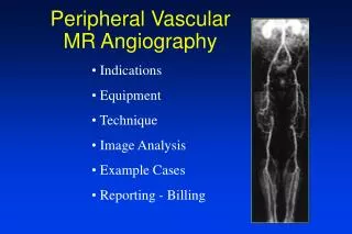
Doppler Ultrasonography • Doppler Ultrasonography • Each arm • Dorsalis pedis • Posterior tibial • Doppler Ultrasonography: • Definition: Doppler ultrasonography is a non-invasive diagnostic procedure that changes sound waves into an image that can be viewed on a monitor. • Purpose: Doppler ultrasonography can detect the direction, velocity, and turbulence of blood flow. It is frequently used to detect problems with heart valves or to measure blood flow through the arteries. • The test is widely used because it is noninvasive, uses no x rays, and gives excellent images. It is harmless, painless, and widely available.
Preparation: There is no special preparation needed for this test. The ultrasound technician may apply a clear gel to the skin in order to help the transducer more freely over the body. • Aftercare: No aftercare is necessary. • Normal results: A Doppler ultrasonography test showing no restricted blood flow is a normal finding. • Abnormal results: Disrupted or obstructed blood flow through the neck arteries may indicate the person is a risk of having a stroke. (Narrowed arterial flow in the legs does not necessarily indicate a risk of stroke.)
Arteriograms & Arteriography • Description .It is invasive procedure involves visualization of arteries, the most common site for an arteriogram is the femoral artery. . Arterial catheters are positioned with a guide wire that is used to advance the catheter to a special location in the arterial tree. . Because the arterial catheter is radiopaque, movement of catheter is noted with fluoroscopy.
Arteriograms & Arteriography . After correct placement is obtained, dye is injected into the catheter to outline a portion of the artery. In this way the circulation in the lower extremity can be visualized. . The catheter can be threaded via the femoral artery into the abdominal aorta to the level of the renal arteries for renal arteriogram. . Other sites for arteriography include brachial artery & carotid artery
Arteriograms & Arteriography • Purposes • Observe blood flow • Detect any lesion • Diagnosed of kidney or liver lesion • Introduction of chemotherapeutic drugs • Introduction of drugs to stop bleeding • Remove atherosclerotic plaques
Arteriograms & Arteriography • Client preparation; . Before .. NPO 6- 8 hrs .. Consent form .. Body weight to determine dose of dye .. Shaving the area . After .. Pressure dressing should not be removed .. Stay supine 4 -6 hrs because of fear from bleeding .. Ice bag may apply to puncture site .. V/S Q 15 min 1st hr then Q2-4 hrs .. Observe pulses distal to arterial puncture