Download
1 / 49
490 likes | 516 Vues
Learn lung volumes, airway pressures, gas exchange, oxygenation, and more for critical care in ICU. Detailed objectives and methods discussed for effective patient care.

E N D
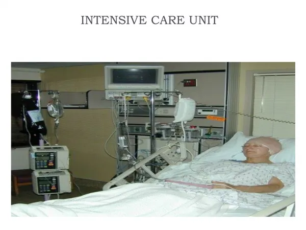
Chapter 14 Respiratory Monitoring in the Intensive Care Unit
Learning Objectives After reading this chapter you will be able to: Identify the methods, normal values, and significance of measuring the following lung volumes in the ICU: tidal volume, rapid-shallow breathing index, vital capacity, functional residual capacity
Learning Objectives (cont’d) Identify the methods, normal values, and significance of measuring the following airway pressures or related indices in the ICU: peak pressure, plateau pressure, compliance, airway resistance, mean airway pressure, maximum inspiratory pressure List the definition, methods of detection, and methods of minimizing auto-PEEP
Learning Objectives (cont’d) Describe value of monitoring pressure, volume and flow waveforms, and pressure volume curves in mechanically ventilated patients Describe methods and significance of measuring the fraction of inspired oxygen and exhaled carbon dioxide in the ICU List the components of oxygen transport and their significance List components of clinical evaluation of oxygenation and their significance
Learning Objectives (cont’d) Explain how these parameters can evaluate tissue oxygen delivery and use: Oxygen delivery and availability Oxygen consumption Mixed venous oxygen tension Venous saturation Arterial to mixed venous oxygen content difference Oxygen extraction ratio Blood lactate
Learning Objectives (cont’d) Describe the value and limitations of pulse oximetry in monitoring oxygenation and oxygen delivery Identify the techniques for monitoring tissue oxygenation and utilization
Overview Monitoring: repeated or continuous observations or measurements of the patient Guide therapeutic interventions Assess interventions Alert clinicians to changes in patient’s condition
Ventilatory Assessment PaCO2: standard for assessing ventilation Changes in metabolism, lung mechanics, ventilatory efficiency, equipment function may precede changes in blood gas Ventilatory parameters monitored Lung volumes and flows Airway pressures Fractional gas concentrations
Lung Volumes and Flows Why? They affect gas exchange They reflect changes in patient’s clinical status They indicate response to therapy They signal problems with patient/ventilator interface Who? Intubated and nonintubated patients
Lung Volumes and Flows (cont’d) What do we measure? VT (5-8 ml/kg IBW) VT<5 ml/kg may indicate respiratory problem Pneumonia, COPD, CHF, ARDS, CNS depression Large VT in metabolic acidosis, sepsis, neurological injury VT = VA + VD VD = 25% to 40% of the VT VD >60% = need ventilatory support
Lung Volumes and Flows (cont’d) High VT ventilation with positive pressure: volutrauma PEEP + smaller VT maintains FRC Discrepancies between set/measured VT Compressible volume of the circuit Inspiratory and expiratory flow sensors Pneumothorax Leaks in the circuit or ETT
Lung Volumes and Flows (cont’d) Spontaneous breathing trial (SBT) failure: VT <300 ml or <4 ml/kg SpO2 <85% to 90% Blood pressure and heart rate change >20% Respiratory rate >35/min Change in mental status Accessory muscle use Diaphoresis
Rapid shallow breathing index (RSBI) RSBI = f (breaths/min)/VT (liters) RSBI >105: prognostic of failure VE = 5 to 6 L/min VE > 10 L/min: weaning not likely successful Lung Volumes and Flows (cont’d)
Vital capacity (VC) 65 to 75 ml/kg IBW FVC <20 ml/kg preoperative: risk of pulmonary complications VC 10 to 15 ml/kg needed for deep breathing and coughing VC >10 to 15 ml/kg for successful weaning and extubation Lung Volumes and Flows (cont’d)
Functional residual capacity (FRC) FRC = 40 ml/kg IBW PEEP and CPAP increase FRC Beneficial in atelectasis and refractory hypoxemia as occurs with ARDS Lung Volumes and Flows (cont’d)
Airway Pressures Important to monitor airway pressures: Need for mechanical ventilation and readiness for weaning Determine site and cause of impedance to mechanical ventilation Evaluate elastic recoil, compliance of thorax Estimate amount of airway pressure transmitted to heart and major vessels Assess respiratory muscle strength
Airway Pressures (cont’d) Peak pressure or PIP Pressure required to overcome opposition to airflow in the lungs Increased resistance Bronchospasm, airway secretions, mucus plugging Decreased compliance (lung or chest wall) Patient-ventilator interface problem
Airway Pressures (cont’d) Plateau pressure Elastic recoil of lung and chest wall Static pressure during period of no gas flow Pressure required to maintain inflation
Airway Pressures (cont’d) Mean airway pressure (Paw) Affected by CPAP, PEEP, inspiratory time (TI), VT, PIP, and rate Paw= [½ (PIP – PEEP) ×(inspiratory time/total cycle time)] + PEEP Impacts oxygenation Caution when Paw >20 cm H2O _ _ _
Airway Pressures (cont’d) Maximum inspiratory pressure (PImax) Influenced by: Respiratory muscle strength Patient effort/ventilatory drive Lung volume Phrenic nerve function Nutritional status Oxygenation/acid-base status Normal PImax: –80 to –100 cm H2O PImax> –30 cm H2O may be useful to predict successful weaning
Airway Pressures (cont’d) Auto-PEEP Total PEEP – set PEEP Reduction of auto-PEEP Bronchodilator therapy Decreased TI (allows more time for exhalation) Reduction of mechanical frequency Reduction of VE is the most effective
Airway Pressures (cont’d) Compliance Dynamic compliance = {Corr VT – [(PIP – EEP) × CF]}/PIP – EEP Static compliance = Corr VT - [(Pplat - EEP) × CF]/Pplat – EEP Normal static compliance in patients receiving mechanical ventilation: 40 to 80 ml/cm H2O Compliance <20 – 25 ml/cm H2O associated with failure to wean
Airway Pressures (cont’d) Airway resistance (Raw) (Ra-w = PIP (cm H2O) – plateau pressure (cm H2O)/flow (L/sec) Normal = 1 to 3 cm H2O/L/sec _ _
Integrating Pressure, Flow, and Volume Evaluating patient/ventilator interface At the patient: use of accessory muscles, color, diaphoresis, heart rate, respiratory rate At the airway: type, size, integrity, stability At the ventilator circuit: leaks, temperature, condensate At the ventilator settings and monitoring panel
Integrating Pressure, Flow, and Volume (cont’d) Monitoring pressure, flow, and volume Graphic display screen Scalar: a single parameter over time Pressure-time waveform Volume-time waveform Flow-time waveform Loop: two parameters in a continuous tracing Pressure-volume loop Flow-volume loop
Integrating Pressure, Flow, and Volume (cont’d) Titrating PEEP and tidal volume with P/V Static pressure-volume curve Super syringe technique Time consuming and cumbersome Useful in acute lung injury Lower inflection point + 2 cm H2O: minimal PEEP Upper inflection point: overdistention
Fractional Gas Concentrations FIO2 Exhaled CO2 Capnometry and capnography Affected by temperature changes, shivering, seizures, trauma, high carbohydrate infusion Measure efficiency of ventilation PETCO2: accurate estimate of PaCO2 CPR effectiveness
Dead Space-Tidal Volume Ratio VD/VT Anatomic dead space: 1 ml/kg IBW Alveolar dead space VD/VT = PaCO2 – PE- CO2 /PaCO2 Normal: 25% to 40% _
Evaluation of Oxygenation Evaluation of oxygen transport Oxygen consumption Oxygen delivery (DO2) Oxygen reserves Oxygen content CaO2 = (Hb × 1.34 × % saturation) + (PaO2 × 0.003) Cardiac output Oxyhemoglobin dissociation curve
Evaluation of Oxygenation (cont’d) Monitoring adequacy of arterial oxygenation Partial pressure of arterial oxygen Should be kept 60 to 80 mm Hg Alveolar-arterial oxygen tension difference P(A-a)O2 PaO2/FIO2 ratio ALI: 200 to 300 ARDS: <200
Monitoring adequacy of arterial oxygenation Oxygen index = Paw × FIO 2 × 100/PaO2 OI >40: mortality >80% Evaluation of Oxygenation (cont’d) _ _
Intrapulmonary shunt (QS/QT) Increased in atelectasis, pneumonia, ARDS, pulmonary edema Pulse oximetry will reveal low SpO2 with elevated FIO2 Co-oximeter useful to determine CaO2 Evaluation of Oxygenation (cont’d) . .
Monitoring Tissue Oxygenation Oxygen delivery (DO2) Cardiac output × CaO2 ×10* Normal: 550 to 650 ml/min/m2 Oxygen consumption (VO2) Fick principle Normal: 100 to 140 ml/min/m2 *Change vol% to ml/L .
Monitoring Tissue Oxygenation (cont’d) Mixed venous oxygen tension PvO2 Normal: 38 to 42 mm Hg Low: inadequate cardiac output, anemia, hypoxia High: poor sampling technique, left-to-right shunt, septic shock, increased cardiac output, cyanide poisoning _
Monitoring Tissue Oxygenation (cont’d) Mixed venous oxygen saturation (SvO2) Fiberoptic reflectance oximetry Decreased: suctioning, shivering, extubation, weaning, PPV _
Monitoring Tissue Oxygenation (cont’d) Arterial-mixed venous oxygen content difference, C(a – v)O2 reflects: Normal: 4 to 6 vol% Increased: low cardiac output, increasing VO2 Decreased: septic shock, increased cardiac output, anemia, left shift ODC _ .
Monitoring Tissue Oxygenation (cont’d) Oxygen extraction ratio [C(a – v)O2/CaO2] Normal: 25% to 30% Increased: low cardiac output, increased VO2, decreased CaO2, Decreased: high cardiac output, sepsis _ .
Monitoring Tissue Oxygenation (cont’d) Blood lactate Anaerobic metabolism = lactic acid production Normal: <1.7 to 2.0 mM/L >3.83 mM/L = 67% mortality >8 mM/L = 90% mortality
Summary Common ventilatory measurements Lung volumes and flows Airway pressures Fractional gas concentrations These measurements allow clinicians to determine the need for mechanical ventilation, monitor the patient, and determine the readiness for weaning