Download

1 / 19
190 likes | 215 Vues
Explore the practical use and key characteristics of fluorescent proteins for advanced live cell microscopy. Discover the diverse applications, properties, and structures of genetically encoded fluorescent proteins enhancing live cell imaging.

E N D
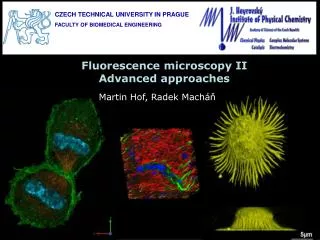
ADVANCED LIGHT MICROSCOPY UNIT Centros Científicos y Tecnológicos Universidad de Barcelona Bellvitge Campus Esther Castaño Benjamín Torrejón torrejonbenja@ccit.ub.edu Casanova Campus Anna Bosch Maria Calvo Elisenda Coll Diagonal Campus Manel Bosch www.ccit.ub.edu
Fluorescent proteins for live cell microscopy Image kindly provided by Dra. Soledad Alcántara
FLUOROPHORES FOR LIFE CELL IMAGING AND SIGNALLING • Cell viability (apoptosis, cell cycle, live/dead…) • 2. General morphology and structure (shape, volume, motility…) • 3. Specific organelles: • a. Nucleus • b. Mitochondrial and mitochondrial membrane potential • c. ER and Golgi • d. Ion physiology (Ca2+, Na+, H+, pH…) • e. Tracers for membrane and cells • 4. Fluorescent proteins (GFP and variants, RPF, dsRed…)
FLUORESCENT PROTEINS • Genetically encoded: fusion with protein of interest (quimera). No microinjection disruption • 2. Fusion protein maintains function and subcellular localization. • 3. Photostable: long period Imaging • 4. High fluorescence yield (bright) • 5. Resistant to photobleaching when illuminated with low intensity • 6. GFP properties have increased biological aplications of live cell imaging and photobleaching. Aequora victora Green Fluorescent Protein Absorption spectra Emision spectra Discosoma striata Red Fluorescent Protein
GFP-like fluorescent proteins structure • 220-240 AA (25 KDa) • Barrel 11 β-sheets and internal disorted helix • Chromophore AA 65-67 in the middle • Barrel is stabilized by noncovalent interactions • High stability to thermal and chemical denaturation and resistence to proteolysis
KEY CHARACTERISTICS FOR FPs PRACTICAL USE 1. Excitation and emission spectra Spectral diversity of available monomeric FPs. Columns show positions of emission maxima and relative brightness of representative monomers
KEY CHARACTERISTICS FOR FPs PRACTICAL USE • Brightness: Quantum yield and extinction coefficient • Quantum yield (Φ) is the emission efficiency of a given fluorophore Loss of energy (heat…). Max value 1 Molar extinction coefficient (ε) is defined as the quantity of light that ca be absorbed by a fluorophore at a given wavelenght and is measured in M-1 cm-1 Brightness = Extinction coefficient x quantum yield • pH stability • pKa is the pH value at which the brightness of fluorescence equals 50% of the maximal brightness measured at an optimal pH
KEY CHARACTERISTICS FOR FPs PRACTICAL USE • Maturation rate • Is the time that takes a FP to become fluorescent (chromophore maturation depends on FP characteristics, oxygen concentration and temperature) • Photostability and undesirable photoconversions • Depends on light source, intensity of light, frequency of light pulses, excitation wavelenghts… • Oligomeric nature and aggregation • Use of fusion proteins is restricted to monomeric FP’s. Dimerization of GFP-like FP at high concentrations can be eliminated by a point mutation (A206K). Engineering a red monomeric FP that wouldn’t aggregate.
APPLICATIONS OF FLUORESCENT PROTEINS • 1. Protein labeling • Cloning a gene of interest in frame with an FP results in a genetic construct that can be transferred into cells and organisms. GFP-like based fusion proteins usually do not affect its proper localization and function. • 2. Photobleaching techniques • Study the mobility of FPs. Fluorescence recovery after photobleaching (FRAP), Fluorescence loss in photobleaching (FLIP)…
APPLICATIONS OF FLUORESCENT PROTEINS 3. Promoters tracking (specific cell and tissue labelling) An FP can highlight promoter activity in a given genetic environment, in particular cells and tissues, in particular time, and in response to an external influence. Schematic representation of multicolor cell labeling using Brainbow technology
APPLICATIONS OF FLUORESCENT PROTEINS • 4. Subcellular localization • An FP-based sensor construct may be thus targeted by an specific AA sequence to various subcellular compartments, enabling organelle visualization, studies of fusion-fission events, and local monitoring of cellular parameters. • Timers • Timer FPs can change fluorescence color with time (related to various maturation rates) and thus allow the determination of their temporal expression in retrospect, ex DsRed-E5 green but later (hours) converts to red. • DNA and RNA labeling • Protein-protein interactions: Förster Resonance Energy Transfer (FRET), Fluorescence correlation spectroscopy (FCS), Fluorescence complementation (split FP’s)…
APPLICATIONS OF FLUORESCENT PROTEINS 8. Photoactivable fluorescent proteins (PAFPs) PAFP fluorescent properties can be turned on by pulse of light of a specific wavelenght (from the dark state or switching from one color to another). Selected irreversibly photoactivatable FPs
REFERENCES Chudakov DM, Matz MV, Lukyanov S, Lukyanov KA. Fluorescent proteins and their applications in imaging living cells and tissues. Physiol Rev. 2010 Jul;90(3):1103-63. Review. PubMed PMID: 20664080. THANKS TO ALL DISCUSSION