Purification and Characterization of High Molecular Weight Dsp Proteins via RP-HPLC and SDS-PAGE
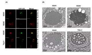
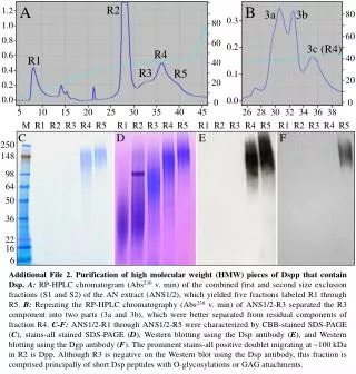
This study outlines the purification process of high molecular weight (HMW) components of Dsp using RP-HPLC. The procedure involved multiple size exclusion fractionations, yielding five distinct fractions (R1-R5) for further analysis. The RP-HPLC chromatography of combined fractions revealed effective separation of components, particularly highlighting the distinct characteristics of each fraction through CBB-stained SDS-PAGE, stains-all, and Western blotting with Dsp and Dgp antibodies. Notably, fraction R2 exhibited a prominent doublet at ~100 kDa, indicative of Dpp, while R3 was primarily composed of short Dsp peptides.

Purification and Characterization of High Molecular Weight Dsp Proteins via RP-HPLC and SDS-PAGE
E N D
Presentation Transcript
A B R2 1.2 3a 3b 80 0.3 80 1.0 60 0.8 60 0.2 3c (R4) R4 0.6 40 R1 40 0.4 R3 R5 0.1 20 20 0.2 0.0 0.0 0 0 5 10 15 20 25 30 35 40 45 26 28 30 32 34 36 38 M R1 R2 R3 R4 R5 R1 R2 R3 R4 R5 R1 R2 R3 R4 R5 R1 R2 R3 R4 R5 C D E F 250 148 98 64 50 36 22 16 6 Additional File 2. Purification of high molecular weight (HMW) pieces of Dspp that contain Dsp.A: RP-HPLC chromatogram (Abs230 v. min) of the combined first and second size exclusion fractions (S1 and S2) of the AN extract (ANS1/2), which yielded five fractions labeled R1 through R5. B: Repeating the RP-HPLC chromatography (Abs230 v. min) of ANS1/2-R3 separated the R3 component into two parts (3a and 3b), which were better separated from residual components of fraction R4. C-F: ANS1/2-R1 through ANS1/2-R5 were characterized by CBB-stained SDS-PAGE (C), stains-all stained SDS-PAGE (D), Western blotting using the Dsp antibody (E), and Western blotting using the Dgp antibody (F). The prominent stains-all positive doublet migrating at ~100 kDa in R2 is Dpp. Although R3 is negative on the Western blot using the Dsp antibody, this fraction is comprised principally of short Dsp peptides with O-glycosylations or GAG attachments.