
Vascular Surgery: Assessment of Arterial Circulation in Limb
E N D
Presentation Transcript
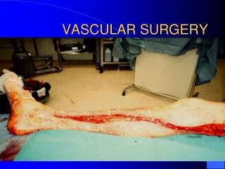
Assessment of arterial circulation in limp. • Inspect: • Color: marble white, blue hue, cyanosed • Vascular angle- Burger's angle: lift leg to white look at what angle the leg is. • Capillary filling time: after lifting the leg but it over the bed and see how long it takes for skin to turn pink. • Venous filling: look for guttering, and angle of venous loss. • Pressure areas: look particularly closely at these, as changes will be first apparent here, trophic, ulceration, gangrenous changes-heel, malleoli, head of 5th metatarsal, tips of toes, between toes, ball of foot.
Assessment of arterial circulation in limp • Palpation: • Temperature: after 5 min exposure to ambiant temp. • Capillary refilling: press tip of toe. • Pulses: fem, dorsalis pedis, posterial tibial artery (medial malleoli), popliteal. • Test muscles/nerves- may be affected by ischaemia. • Auscultation: • over all major arteries • Blood pressure in both armes.
ISCHEMIA It the condition of inadequate blood supply to an area of tissue producing harmful effect to its function &nutrition
Acute Ischemia It the condition of inadequate blood supply to an area of tissue producing harmful effect to its function &nutrition of less than 2 weeks duration
Acute Ischemia ( etiology) • Embolism • Thrombosis • Others • Acute arterial trauma • Dissecting aortic aneurysm • Compartmental syndrome • External compression • Poploteal entrapment • Cystic adventational disease
Management of acute ischemia • Investigations • Urea, electrolytes, BSL • ECG, chest x ray • Initial treatment • Rehydration • I V analgesia • Heparinization
Acute LL ischemia Less sever LL ischemia (previously ischemic limb) Sever LL ischemia (previously normal limb) Arterio-graphy must be done to determine site, size& extent of thrombus occlusion Fogarty embolectomy
Sever LL ischemia (previously normal limb) Femoral embolectomy (assess degree of inflow) Poor inflow Good inflow Distal embolectomy Perform proximal Iliac embolectomy Perform intra-operative arteriogram To assess efficiency Poor inflow No occlusion Residual thrombus Proximal vascular reconstruction Close arteiotomy& Perform fasciotomy Thrombolysis
Less sever LL ischemia (previously ischemic limb) Arterio-graphy Thrombolysis After the underlying Cause is detected If thrombo-lysis are contraindicated By pass procedure to site of occlusion or angioplasty
Thrombo-lysis • This depend on per-cutaneous delivery of thrombo-lytic drugs within the thrombus • to dissolve it by intra-arterial catheter placed within the thrombus • Most centers in UK have limited the technique for thrombus less than 30 days
Thrombo-lysis • Drugs • Streptokinase • Urokinase • Recombinant TPA • Technique • Local low dose • Pulse spray technique • High dose bolus technique • Check angio-graphy is done every 8-9 hous and catheter tip repositioned as necessary • Success is 60- 70 % with careful selection
Thrombo-lysis • Complications • Mortality 1-2 % • Major bleeding 10 % • Minor bleeding 25 % • Stroke • Embolization • Contraindications • Bleeding diathesis • Long term anticoagulant • Stroke • Old age > 75 Y • Peptic ulcer
Chronic LL ischemia (Risk factors) Sedentary life Obesity Age & sex Risk factors of LL ischemia Hyper- lipidaemia Diabetes Hypertension Smoking
Chronic LL ischemia (Etiology) Age above 45 Y Age below 45 years Atherosclerosis is the commonest cause Diabetics Non Diabetics In males smokers In both In female Pre-senile atherosclerosis Raynaud’s disease Burger’s disease Arteritis
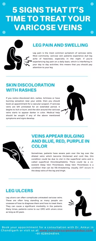
Chronic LL ischemia (Clinically) Press & See How Color Fade (pre-gangrene) & (gangrene) Pain Sensation Hotness Color Function Pre-gangrene (Nutritional) Gangrene
Chronic LL ischemia (DD) • Nerve compression (Sciatica) • Veins ( CVI and DVT) • Joints (arthropathy) • Muscle ( myopathy) • Bone pains • Superficial lesions in skin& Sc tissue
Where is the site of occlusion ? Aortoiliac pattern • Claudication gluteal region • Wasting of thigh muscles • Lost or weak femoral pulses • Impotence if bilateral Inguinal ligament Femoro popliteal pattern Adductor hiatus • Claudication calf • Lost or weak popliteal pulse • Beurger’s sign (pallor on elevation and rubor on dependency) Distal circulation pattern
Chronic LL ischemia(thrombosis) Le Riche syndrome • Claudication in gluteal region • Wasting of thigh muscles • Lost or weak femoral pulses • Impotence
Claudication foot • Lost or weak dorsalis pedis and or posterior tibial • Beuger’s sign • Nutritional changes (10 items) Aortoiliac pattern Inguinal ligament Femoropopliteal pattern • Skin, • skin appendages, • subcutaneous fat, • muscles, • ulcers, • gangrene, • delayed venous filling, • coldness, • motor and • sensory changes Adductor hiatus Distal circulation pattern
Vascular lab • Segmental limb pressure • Ankle- Brachial Index • Normal > 1 • Intermittent claudication 0.5- 0.9 • No healing < 0.5 • Rest pain 0.4
Vascular lab • Toe- brachial Index • Normal 0.8- 0.9 • Caludicate 0.35 • Rest pain 0.1 • Toe pressures • Normal 90 – 100 mmHg • CLI < 30 mmHg • Exercise tests
Non Invasive • Doppler U/S • Duplex U/S • Plethysmography • Isotope blood flow • Trans-cutaneous oxygen tension
Invasive (Arteriography) It is the gold stander of arterial tree Methods • Directly trans-femoral if pulse is palpable • Seldinger approach • Digital sub-straction angiography contrast material injected I.V in large volumes or IA. In small tiny volumes
Invasive (Aorto-graphy) »Translumbar if both F pulses are not felt »Transfemoral aortography if one F pulse is felt »Transbrachial if the entire distal aorta is occluded »Digital sub-straction angiography
Invasive (Arteriography) Potential complications include • Contrast-related • Anaphylactic reaction • Toxic reactions • Deterioration in renal function • Technique-related • Haematoma • Arterial spasm • Sub-intimal dissection • False aneurysm • Arteriovenous fistula • Embolisation • Infection
New imaging modalites • MR angiography (is now providing the most sensitive test for identifying tibial vessels) • CT angiography which is articularly useful for the assessment of aneurysmal disease • Angioscopy • Intravascular ultrasonography
Treatment of chronic LL ischemia I - Risk factor reduction • Stop smoking - arrests disease progression • Lipid-lowering drugs • Anti-platelet medication • Good diabetic control if appropriate II- Regular exercise • as part of supervised exercise program • Lose weight
Treatment of chronic LL ischemia III – Pharmaco-therapy • Vasodilator drugs with small benefits • Naftidrofyl oxalate, Praxiline • Pentoxyifyllin, Trental 400 • Prostacyclin • Vasodilator drugs with minimal benefits • Antiplatlets ( aspirin) • Prostaglandins • Ca channel blockers
Endo vascular surgery Basic principles • The symptoms should be life-style limiting • Co-management of underlying conditions likely to limit safety or success (smoking, heart failure etc) • Proximal disease should be managed before distal ones • Localized (<10 cm) non-ulcerating lesion is an ideal lesion
Endo vascular surgery Basic techniques •Balloon dilatation •Stents • Atherectomy devices • Lasers • Vibrating and rotating wires
Percutaneous transluminal angioplasty • Angioplasty of the aorto-iliac segment has a 90% 5 year patency • Angioplasty of the infra-inguinal vessels has a 70% 5 year patency • Best results seen with short segment stenoses less than 2 cm long Complications occur in less than 2% of patients • Wound haematoma • Acute thrombosis • Distal embolisation • Arterial wall rupture
Percutaneous transluminal angioplasty with stents Use of stents • Most are used to correct inadequacies or complications of PTA • To avoid re-stenosis which occurs within 90 days of PTA • When there is significant residual gradient or stenosis following PTA • When there is acute occlusions during PTA • When there is dissection longer than PTA site
Surgical treatment of claudication and rest pain • Indications for surgery: • claudication is a relative indication. • rest pain if fit for operation. • Ischaemic ulceration that does not respone to conservative management. • acute occlusion. • After decision on surgery is done do arteriography: site, type of operation and if technically possible
Surgical treatment Direct arterial surgery Indirect arterial surgery Sympathectomy Bypass Thromb endarterectomy Amputation