Download
1 / 1
10 likes | 139 Vues
Binding of anti-OPN mAbs to Osteopontin: Epitope Recognition Under Various Conditions Monica Kadia, Nady Hin, Tanya Gordonov, Yacov Ron, Esben S ø rensen, Larry Steinman and David T. Denhardt.

E N D
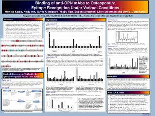
Binding of anti-OPN mAbs to Osteopontin: Epitope Recognition Under Various Conditions Monica Kadia, Nady Hin, Tanya Gordonov, Yacov Ron, Esben Sørensen, Larry Steinman and David T. Denhardt Rutgers University (MK, NH, TG, DTD), RMDNJ-UMDNJ (YR), Aarhus University (ES) and Stanford University (LS) Introduction Experiment 4 Objective: Test the variation of binding using two different buffers. SBF (simulated body fluid – mimics the ionic composition of plasma) vs. PBS (phosphate-buffered saline). Procedure: The procedure for using SBF differed from the PBS in that the wash steps all used SBF instead of PBS. Additionally, the biotinylated peptides as well as the primary antibodies were all diluted in SBF as opposed to PBS. Results: We found that, relative to PBS, SBF reduced the binding strength of the mAbs to the “sticky” peptides. The sticky peptides are peptides 11 and 15/16. Experiment 5 Objective: To determine if binding of some mAbs to their presumed cognate epitope can be epitope bind to a nonsticky peptide in the fluorescence assay and not just the sticky peptides. Results: Some mAbs exhibited a strong binding comparable to the positive control to one or more peptides other than the sticky peptides 11 and 15/16 – e.g. 7B4 below. Presumably 7B4 is binding to related sequences in this region of OPN. Experiments • Osteopontin (OPN) is a phosphoglycoprotein that is found in various tissues and body fluids (1). A majority of its functions are affected by its post translational modifications including phosphorylation and glycosylation (2). Anti-OPN monoclonal antibodies have been isolated and characterized by our laboratory (3, 4). Among other findings is evidence for an association of the C-terminal region of the protein with the central RGD region. OPN is found in high levels in patients with disseminated carcinomas. Its expression is enhanced in activated T-lymphocytes in response to certain infections . OPN has various functions, generally acting through certain integrins and/or CD44 variants to promote the survival of cells that have been compromised in various ways. • This research was supported in part by funds from the National Multiple Sclerosis Society, the National Institutes of Health and a donation from Mrs. J. G. Harrar. Experiment 1 Objective: To identify the epitope recognized by each anti-OPN mAb by determining the sequences in the OPN molecule to which the mAb could bind. Fig. 1 shows one set of peptides; a second set comprises sequences bracketing the overlapping domains. Procedure: Using HRP as the secondary antibody and SBF (Simulated Body Fluid) as the buffer, we tested each of the putative anti-OPN monoclonal antibodies against the biotinylated OPN peptides as illustrated in fig. 2. Results: We were able to identify specific mAbs that bound reproducibly and uniquely to a specific region in the OPN molecule. Experiment 2 Objective: Test the variation of binding using two different secondary antibodies, either tagged with a fluorescent label or with HRP. Procedure: The procedure for the fluorescent secondary differed from the HRP secondary in that the fluorescent tag required a black high binding capacity neutravidin-coasted plate as opposed to a clear high binding capacity neutravidin-coated plate; further, the HRP required the use of ABTS to quantify the peroxidase activity. Also, the machines used to read the plates were different. Results: We were able to show that HRP reduces the background and amplifies the weaker signals found in the fluorescent assays. We discovered that most of the putative anti-OPN mAbs bound non-specifically, presumably via the Fc portion, to two “sticky” peptides. Experiment 3 Objective: To demonstrate that a few of the mAbs bind to two adjacent (overlapping) peptides. Results: We found two mAbs that bound to adjacent overlapping regions thus identifying a more precise location of the epitope. Figure 6: This figure shows the suppression of “sticky” peptide binding in SBF buffer. Figure 3. This figure shows mAbs 50H9, 2C5, and 67F7 binding to peptide 2, overlap 9/10, and peptide 19 respectively. All three were tested together; when tested separately each bound only to the indicated peptide. 2A1 (06) is the positive control. Figure 7. This figure shows 7B4 binding to peptide 19 as well as overlap 17/18 (done in SBF). The sequence SHE is found twice in peptide 17/18 and once in 19, which also contains the sequence SKE; 17/18 also contains and SRE sequence. Fig. 1: Sequence of human OPN showing 20 overlapping peptides. Red: Phosphorylated residues; blue: O-glycosylated residues (1). A second set of peptides encompasses the overlapping regions along with adjacent residues. Goals of this research: To identify the epitopes recognized by anti-OPN mAbs Discussion Approach In this project we investigated several conditions in order to see what effect the variations had on the binding of the mAb to the peptides. Binding reactions were done either in PBS or SBF (simulated body fluid). The secondary antibodies was goat anti-mouse IgG coupled either with an AlexaFluor fluorescent tag or with HRP (horse radish peroxidase). Method • Using HRP-conjugated goat anti-mouse IgG as the secondary antibody in place of the IgG secondary antibody tagged with a fluorescent probe resulted in amplification of signals from the actual epitope and a reduction in background binding. • Some monoclonal antibodies showed strong binding to a particular peptide as well as the adjacent overlapping region. This allows us to localize the true epitope more accurately. • Using SBF instead of PBS reduced the binding of the “sticky” peptides to the antibody. • Although the fluorescence assay showed weak binding in general except in the cases of the “sticky” peptides, there were some instances where strong binding to an epitope did occur. Figure 4b. 42B12 binding quantified using the fluorescent secondary antibody. The binding to13/14 was much reduced in comparison with the “sticky” peptides 11 and 15/16. We believe this non-specific binding is mediated by the Fc fragment. Figure 4a. 42B12 binding quantified using HRP. The binding to the “real” epitope 13/14 was increased relative to the “sticky” peptides 11 and 15/16 (to which many of the mAbs bound including mouse IgG itself). 2A1 is always the positive control. BIBLIOGRAPHY • Sodek J, Ganss B, McKee MD. (2000) Osteopontin. Crit Rev Oral Biol Med. 11:279-303. • Christensen B, Nielsen MS, Haselmann KF, Petersen TE, Sorensen ES. (2005) Post-translationally modified residues of native human osteopontin are located in clusters: identification of 36 phosphorylation and five O-glycosylation sites and their biological implications. Biochem J. 390:285-92. • Kazanecki CC, Uzwiak DJ, Denhardt DT. (2007) Control of osteopontin signaling and function by post-translational phosphorylation and protein folding. J Cell Biochem. 102:912-24. • 4. Kazanecki CC, Kowalski AJ, Ding T, Rittling SR, Denhardt DT. (2007) Characterization of anti-osteopontin monoclonal antibodies: Binding sensitivity to post-translational modifications. J Cell Biochem. 102:925-35. Fig. 5: This figure (generated using SBF and HRP) shows the two mAbs 10H4cl5 and 1H3F7 binding to peptides 13/14 and 14 and to 15 and 15/16 respectively. Binding to other peptides was much lower. This is strong evidence that the actual epitope for 10H4cl5 is contained in the sequence DLNAPSDW and for 1H3F7 is within THSHKQS. We also found that 2A1 bound to peptides 13 and 13/14, which contain the IPVA sequence previously implicated as the critical epitope (4). Fig. 2: Cartoon illustrating the basic outline of the protocol used.