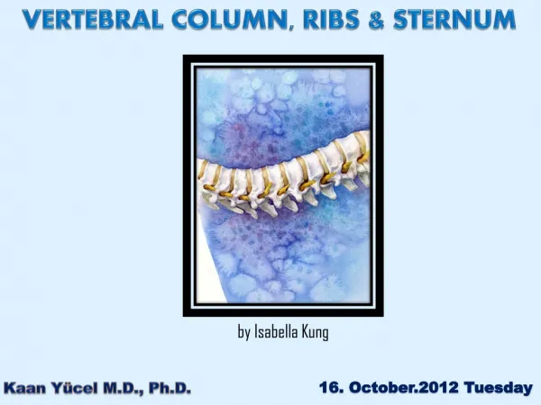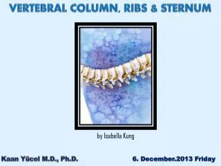
VERTEBRAL COLUMN RIBS STERNUM
VERTEBRAL COLUMN RIBS STERNUM . Yeditepe University Medical School Department of Anatomy. Kaan Yücel M.D., Ph.D. 2. November.2011 Tuesday. Vertebrae + intervertebtal (IV) discs = Vertebral column Spine Omurga Wirbelsäule العمود الفقري Uti

VERTEBRAL COLUMN RIBS STERNUM
E N D
Presentation Transcript
VERTEBRAL COLUMN RIBS STERNUM Yeditepe University Medical School Department of Anatomy Kaan Yücel M.D., Ph.D. 2. November.2011 Tuesday
Vertebrae +intervertebtal (IV) discs= Vertebral column Spine Omurga Wirbelsäule العمود الفقري Uti Skeleton of the neck & back Main part of the axial skeleton
Vertebral column Extends from the cranium (skull) to the apex of the coccyx. In the adult it is 72-75 cm long. ¼ formed by the intervertebral (IV) discs IV discs separate and bind the vertebrae together.
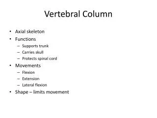
Protectsthe spinal cord and spinal nerves. • Supportsthe weight of the body superior to the level of the pelvis. • Provides a partly rigid and flexible axis for the body and an extended base on which the head is placed and pivots. • Plays an important role in posture and locomotion • (the movement from one place to another). Vertebral column
VERTEBRAE • The vertebral column is flexible because it consists of many relatively small bones, called vertebrae(singular = vertebra), that are separated by resilient intervertebral (IV) discs.
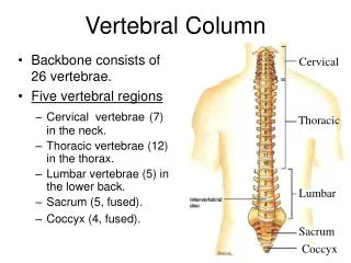
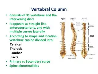
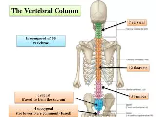
The vertebral column in an adult typically consists of • 33 vertebrae arranged in 5 regions: • 7 cervical • 12 thoracic • 5 lumbar • 5 sacral • 4 coccygeal
Significant motion occurs only between the 25 superior vertebrae. • Of the 9 inferior vertebrae, the 5 sacral vertebrae are fused in adults to form the sacrum • After ~ 30, the 4 coccygeal vertebrae fuse to form the coccyx.
The vertebrae gradually become larger as the vertebral column descends to the sacrum and then become progressively smaller toward the apex of the coccyx.
The 25 cervical, thoracic, lumbar, and first sacral vertebrae also articulate at synovial zygapophysial joints, which facilitate and control the vertebral column's flexibility. • Although the movement between two adjacent vertebrae is small, in aggregate the vertebrae and IV discs uniting them form a remarkably flexible yet rigid column that protects the spinal cord they surround.
Curvatures in the Vertebral Column There are four natural curves in a healthy spine. 1. The neck or cervical spine, curves gently inward (lordosis) 2. The mid back, or thoracic spine, is curved outward (kyphosis) 3. The low back, or lumbar spine, also curves inward (lordosis) 4. Pelvic (Sacral) curvature
Structure and Function of Vertebrae • Vertebrae vary in size and other characteristics from one region of the vertebral column to another, and to a lesser degree within each region; however, their basic structure is the same. • A typical vertebra consists of a • Vertebral body • Vertebral arch • 7 processes
Vertebral body • Massive • Roughly cylindrical • Anterior part of the bone • Gives strength to the vertebral column and supports body weight. • The size of the vertebral bodies the column descends • most markedly from T4 inferiorly • As each bears progressively greater body weight.
Vertebral arch • Posterior to the vertebral body • Consists of two (right and left)pedicles& laminae.
Pedicles • Short, strong cylindrical processes • Project posteriorly from the vertebral body to meet two broad, flat plates of bone, called laminae, which unite in the midline. • The vertebral arch and the posterior surface of the vertebral body • form • the walls of the vertebral foramen
The succession of vertebral foramina • in the articulated vertebral column • forms • the vertebral canal (spinal canal) • Spinal canal • Contains the spinal cord and the roots of the spinal nerves that emerge from it, along with the membranes (meninges), fat, and vessels that surround and serve them.
Vertebral notches(Incisura vertebralis) • Indentations observed in lateral views of the vertebrae • Superior and inferior to each pedicle • Between the superiorand inferior articular processes posteriorly • Between the corresponding projections of the body anteriorly.
The superior and inferior vertebral notchesof adjacent vertebrae and the IV discs form intervertebral foramina • Intervertebral foramina • Spinal (posterior root) ganglia are located • Spinal nerves emerge from the vertebral column with their accompanying vessels through these foramina.
7 processesarise from the vertebral arch of a typical vertebra: • 1 median spinous process projects posteriorly from the vertebral arch at the junction of the laminae. • 2 transverse processes project posterolaterally from the junctions of the pedicles and laminae. • 4 articular processes (G. zygapophyses)—2 superior and 2 inferior—also arise from the junctions of the pedicles and laminae, each bearing an articular surface (facet).
1 median spinous • 2 transverse processes • 4 articular processes (G. zygapophyses)
The spinous and transverse processes provide attachment for deep back muscles and serve as levers, facilitating the muscles that fix or change the position of the vertebrae.
The articular processes are in apposition with corresponding processes of vertebrae adjacent (superior and inferior) to them, forming zygapophysial (facet) joints. • Through their participation in these joints, these processes determine the types of movement permitted and restricted between the adjacent vertebrae of each region.
The articular processes also assist in keeping adjacent vertebrae aligned, particularly preventing one vertebra from slipping • anteriorly on the vertebra below.
Generally, the articular processes bear weight only temporarily, as when one rises from the flexed position, and unilaterally when the cervical vertebrae are laterally flexed to their limit. • However, the inferior articular processes of the L5 vertebra bear weight even in the erect posture.
Regional Characteristics of Vertebrae • Each of the 33 vertebrae is unique. However, most of the vertebrae demonstrate characteristic features identifying them as belonging to one of the 5 regions of the vertebral column (e.g., vertebrae having foramina in their transverse processes are cervical vertebrae).
In addition, certain individual vertebrae have distinguishing features; the C7 vertebra, for example, has the longest spinous process. It forms a prominence under the skin at the back of the neck, especially when the neck is flexed.
In each region, thearticular facets are oriented on the articular processes of the vertebrae in a characteristic direction that determines the type of movement permitted between the adjacent vertebrae and, in aggregate, for the region. • For example, the articular facets of thoracic vertebrae are nearly vertical, and together define an arc centered in the IV disc; this arrangement permits rotation and lateral flexion of the vertebral column in this region.
Regional variations in the size and shape of the vertebral canal accommodate the varying thickness of the spinal cord.
CERVICAL VERTEBRAE Form the skeleton of the neck. The smallest of the 24 movable vertebrae Located between the cranium and the thoracic vertebrae.
Their smaller size reflects the fact that they bear less weight than do the larger inferior vertebrae. The relative thickness of the discs, the nearly horizontal orientation of the articular facets, and the small amount of surrounding body mass give the cervical region the greatest range and variety of movement of all the vertebral regions.
The most distinctive feature of each cervical vertebra is the oval foramen transversarium(transverse foramen) in the transverse process. The vertebral arteries and their accompanying veins pass through the transverse foramina, except those in C7, which transmit only small accessory veins.
The transverse processes of cervical vertebraeend laterally in 2 projections: an anterior tubercleand a posterior tubercle. The tubercles provide attachment for a laterally placed group of cervical muscles. The anterior rami of the cervical spinal nerves course initially on the transverse processes in grooves for spinal nerves between the tubercles.
The anterior tubercles of vertebra C6are called carotid tubercles (Chassaignac tubercles) because the common carotid arteries may be compressed here, in the groove between the tubercle and body, to control bleeding from these vessels. Bleeding may continue because of the carotid's multiple anastomoses of distal branches with adjacent and contralateral branches, but at a slower rate.
Vertebrae C3-C7are the typical cervical vertebrae. Large vertebral foramina to accommodate the cervical enlargement of the spinal cord as a consequence of this region's role in the innervation of the upper limbs. The adjacent cervical vertebrae articulate in a way that permits free flexion and extension and some lateral flexion but restricted rotation..
The elevated superolateral margin uncus of the body (uncinate process) Spinous processes of the C3-C6 vertebrae Short & usually bifid in white people, especially males but usually not as commonly in people of African descent or in females.
C7 is a prominent vertebrathat is characterized by a long spinous process. • prominent process • C7= vertebra prominens • Most prominent spinous process in 70% of people
Atlas (C1) 2 superior-most cervical vertebrae are atypical. Unique in that it has neither a body nor a spinous process. This ring-shaped bone has paired lateral masses that serve the place of a body by bearing the weight of the globe-like cranium in a manner similar to the way that Atlas of Greek mythology bore the weight of the world on his shoulders.
The transverse processes of the atlas arise from the lateral masses, causing them to be more laterally placed than those of the inferior vertebrae. This feature makes the atlas the widest of the cervical vertebrae, thus providing increased leverage for attached muscles. The kidney-shaped, concave superior articular surfaces of the lateral masses articulate with occipital condyles.
Anterior and posterior arches, each of which bears a tubercle in the center of its external aspect, extend between the lateral masses, forming a complete ring. The posterior arch, corresponds to the lamina of a typical vertebra, has a wide groove for the vertebral artery on its superior surface. C1 nerve also runs in this groove.
Vertebra C2, also called the axis, is the strongest of the cervical vertebrae. C1, carrying the cranium, rotates on C2 (e.g., when a person turns the head to indicate “no”). Axis (C2)
The axis has two large, flat bearing surfaces, the superior articular facets, on which the atlas rotates. The distinguishing feature of C2 is the blunt tooth-like dens Both the dens (G. tooth) and the spinal cord inside its coverings (meninges) are encircled by the atlas. The dens lies anterior to the spinal cord and serves as the pivot about which the rotation of the head occurs.
The dens is held in position against the posterior aspect of the anterior arch of the atlas by the transverse ligament of the atlas. Extends from one lateral mass of the atlas to the other, passing between the dens and spinal cord, forming the posterior wall of the “socket” that receives the dens. Prevents posterior (horizontal) displacement of the dens and anterior displacement of the atlas.
C2 has a large bifid spinous process that can be felt deep in the nuchal groove, the superficial vertical groove at the back of the neck.
THORACIC VERTEBRAE Located in the upper back Provide attachment for the ribs. Primary characteristic features of thoracic vertebrae are the costal facets for articulation with ribs.
The middle 4 thoracic vertebrae (T5-T8) demonstrate all the features typical of thoracic vertebrae. • The articular processes of thoracic vertebrae extend vertically with paired, nearly coronally oriented articular facets that define an arc centered in the IV disc. • This arc permits rotation and some lateral flexion of the vertebral column in this region. In fact, the greatest degree of rotation is permitted here.
The T1-T4 vertebrae share some features of cervical vertebrae. T1 is atypical of thoracic vertebrae in that it has a long, almost horizontal spinous process that may be nearly as prominent as that of the vertebra prominens. T1 also has a complete costal facet on the superior edge of its body for the 1st rib and a demifacet on its inferior edge that contributes to the articular surface for the 2nd rib.