Download
1 / 39
400 likes | 666 Vues
Mechanisms of DNA damage-Induced Cell Death. Radiobiology 2012. Radiation-Induced Cellular Damage. Direct DNA damage – easy to recognize that one target must be repaired Membrane damage – Signaling through ceramide/Growth Factor receptors?

E N D
Mechanisms of DNA damage-Induced Cell Death Radiobiology 2012
Radiation-Induced Cellular Damage • Direct DNA damage – easy to recognize that one target must be repaired • Membrane damage – Signaling through ceramide/Growth Factor receptors? • Postmitotic (secondary) DNA damage - due to loss or alteration in genetic material • Bystander Damage – through soluble factors?
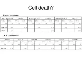
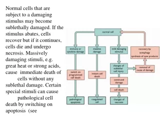
Characterization of Cellular death (in vitro) • Biochemical death – trypan blue positive • Cells unable to exclude dye (loss of membrane potential or membrane) • Reproductive death – colony formation • cells unable to form a colony (may sit very happily on the dish)
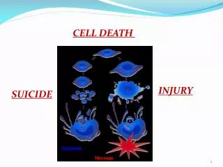
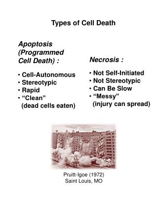
Classification of death processes • Programmed cell death (classical apoptosis and autophagic) • Requires energy, activation of caspases (proteolysis) and nucleases (chromatin fragment) • Necrotic death • Loss of energy leads to ion imbalance across plama membrane (Na/K ATPase) • Mitotic death • Acute mitotic catastrophe, or loss of essential genetic material
Tumor cell reproductive death(Clonogenicdeath) Fraction that Apoptose + Fraction that undergo autophagy + Fraction that undergo mitotic catastrophy + Fraction that enter permanent senescence + Fraction that suffer late mitotic death + Fraction that get lethal bystander damage Fraction of Cells Unable to Grow =
Dose and time-dependent death Shinomiya JCMM 2002
Radiation-Induced Apoptosis • 1992 Lowe showed oncogenically transformed mouse cells apoptosed in response to DNA damage in a p53-dependent manner • Apoptotic p53 effector genes identified: bax, PUMA, noxa, perp (and others) • However, p53 sensitizes to hypoxia without transactivation
Apoptotically sensitive Tumor Cells show clonogenicsensitivity
Apoptotic Cascades • Receptor Mediated • Fas/TNFR family, FADD, Caspase 8, effector caspases • Mitochondrially Mediated • Cytochrome C, APAF-1, Caspase 9, effector caspases • Crosstalk in pathways at C8-Bid acitvation
Bcl2 identified as oncogene because of translocation caused activation Large survival benefit in hematologic malignancies, less significant in solid tumors
BH3-only act as trigger for the apoptotic signal
Mitochondria as Key integrator of Apoptotic signals
Radiation-Induced Apoptotic Signals Belka IJROBP (2004)
Receptor-Mediated Apoptotic Signals Belka IJROBP (2004)
Radiation apoptosis is cell type dependent upon either p53 or ceramide Kolesnick and Fuchs 2002 Science
Apoptotic Modulators as Radiosensizers Belka IJROBP (2004)
In vivo detection of apoptosis Verheig Can Met Rev 2008
In vivo detection of apoptosis using 99T Annexin V Verheig Can Met Rev 2008
Autophagic Death • Autohagy is active “self eating” • Process for degradation of cellular constituents that do not fit in the proteasome (Aggregates or organelles) • Tagging of proteins through ATG5-ATG12 system • Beclin-1 is a haploinsufficient tumor suppressor gene
Autophagic machinery Klionsky Nature 2008
Radiation Senescence • First described in normal cells • Later determined to occur in tumor cells • Senescence-associated B-galactosidase as a marker • p53 and p21 as major contributors
Doxirubicin Treated Colon Cancer Cells Can be Sorted by Proliferation status Non-proliferating cells show Senescent morphology Non-proliferating cells do not Form Colonies Roninson CR 2003)
Mitotic catastrophe • Caused by a combination of deficient cell cycle checkpoint and cellular damage at mitosis • Has characteristics of apoptosis (capase 2 activation) • Thought to prevent aneuploidy, so loss could contribute to tumor progression
Characteristics of Mitotic Catastrophe Mitotic catastrophy induced by chk2 inhibitor (debromohymenialdesine). Castedo Kroemer Onc (2004)
Radiation-Induced Chromosome Aberrations • Exact mechanisms not established, but involves DNA damage and Misrepair • ATM fragile chromosomes and radiosensitivity • Multiple lesions needed for complex aberrations – the D2 component to survival curves • Linear relationship between “lethal” lesions and survival
DSB vs Telomere Purdy CB 2004
Defective DSB Repair Causes Cellular &Clinical Radiation Hypersensitivity From: Hall, “Radiobiology for the Radiologist” From: Hall and Giaccia, “Radiobiology for the Radiologist”
Radiation-Induced aberrations In metaphase spreads
FISH to Identify Aberrations Lymphocytes exposed to 4 Gy Nataragan MR 2003
Asymmetric chromosome aberrations from DSBs • Occur primarily in unreplicated cells (G1) • Can result in direct loss of genetic material • Acentric fragment, internal deletion • Can lead to future generation events • Dicentric bridge, breakage, fusion, bridge • G2 breaks lead to chromatid events, and often one daughter cell has complete DNA content
Resolution of Dicentric Bridge Shimizu ECR 2005
Linear Quadratic relation between Radiation dose and Aberrations
Two linear quadratics make a straight line Survival and asymmetric chromosome lesions are linearly related. Cornforth and Bedford RadRes1987
Bystander Role in Radiation Induced Cellular Death • Photon does not pass through the cell, but there is still a biologic effect • Due to secreted substance (paracrine), or one passed through gap junctions • Can lead to late effect mutation or even cell death • Not really dose-dependent
Clonal effects “Bystander effects” Mothersill and Seymour 2004
Summary • Radiation can induce “death” through a number of established pathways • Most significant fraction is due aberrations in solid tumors • Oncogenic transformation can alter inherent sensitivity to PCD (more myc, less bcl2) • Radiation sensitizers very difficult to achieve because they have to be specific for tumor cells