Download

1 / 23
230 likes | 252 Vues
This review provides knowledge of the anatomy underlying carpal tunnel syndrome, a common workplace injury resulting from compression of the median nerve. Surgical treatment involves decompression of the carpal tunnel. The article discusses the bones of the upper extremity, carpal bones, movements of the forearm and hand, muscles involved in flexion and extension, and the innervation of the upper extremity.

E N D
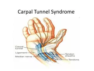
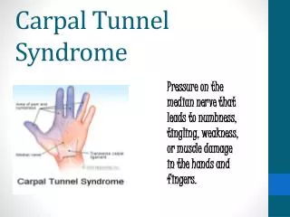
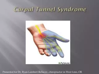
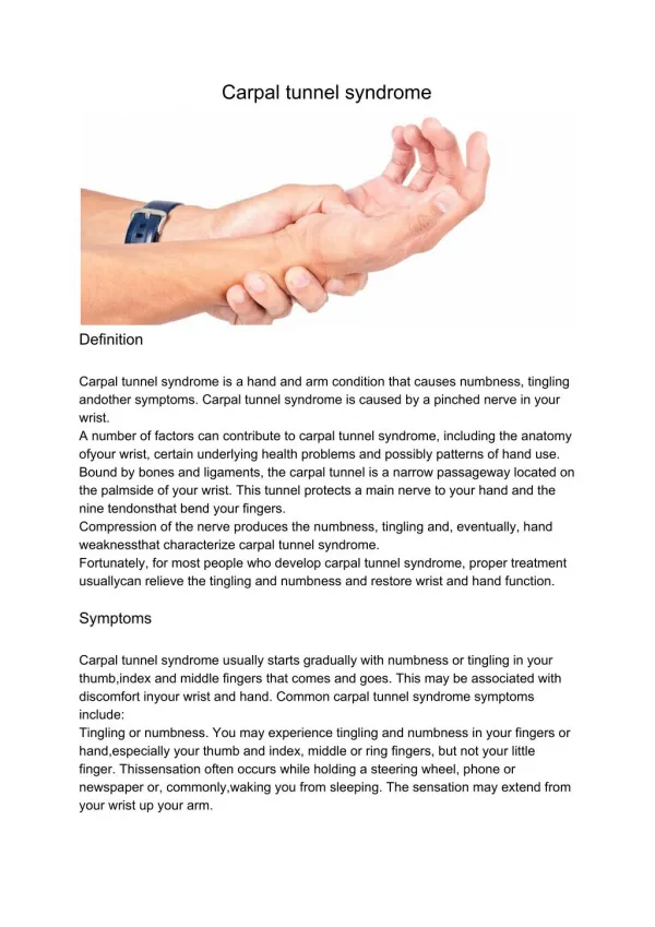
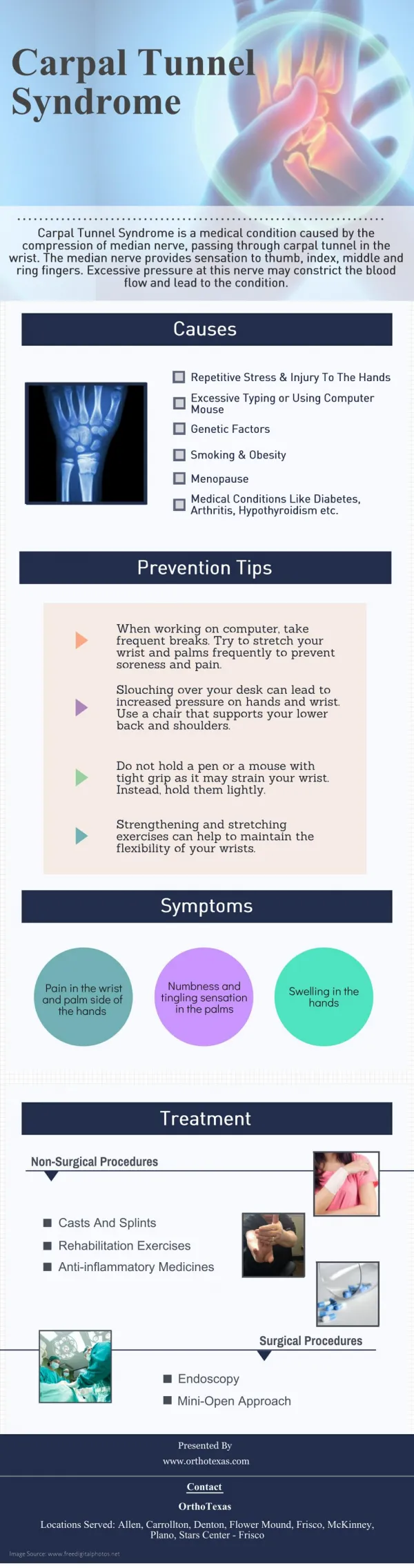
CARPAL TUNNEL SYNDROME Carpal tunnel syndrome is one of the most common workplace injuries. It results from compression of the median nerve as it passes into the hand and can be treated surgically by opening the carpal tunnel (decompression). Knowledge of the anatomy of the upper extremity and hand is essential in the diagnosis and treatment of carpal tunnel syndrome. MEDIAN NERVE
BONES OF UPPER EXTREMITY Clavicle Scapula Humerus Radius Ulna Carpal bones Metacarpals Phalanges CARPAL BONES ARE LOCATED IN THE PROXIMAL PART OF THE HAND
AP RADIOGRAPH OF RIGHT HAND Distal phalanges Middle phalanges Proximal phalanges Metacarpal bones Sesamoid bones Hook of hamate CARPAL BONES Wrist Joint Styloid process of Ulna Styloid process of Radius
ANTERIOR VIEW OF LEFT HAND CARPAL BONES FORM A 'TUNNEL' FOR PASSAGE OF STRUCTURES INTO THE HAND TWO ROWS OF BONES: Order (lateral to medial) Proximal Row - Scaphoid, Lunate, Triquetrum, Pisiform Distal Row - Trapezium, Trapezoid, Capitate, Hamate Capitate Hamate Trapezoid THUMB Trapezium Hook of Hamate Pisiform Triquetrum Lunate Scaphoid MNEMONIC: SOME LOVERS TRY POSITIONS THAT THEY CAN'T HANDLE
AP RADIOGRAPH OF WRIST OF LEFT HAND Hook of Hamate THUMB Hamate First Metacarpal Capitate Trapezium Triquetrum Trapezoid Pisiform Scaphoid Lunate Radial Styloid Process Ulnar Styloid Process Radius Ulna
MOVEMENTS OF FOREARM, HAND EXTENSION/FLEXION SUPINATION/PRONATION EXTENSION AT WRIST, FINGERS SUPINATION = PALM UP (FORWARD) PRONATION= PALM DOWN (BACK) FLEXION AT WRIST, FINGERS
MANY MUSCLES THAT MOVE THE HAND AND FINGERS ARE LOCATED IN THE FOREARM POSTERIOR FOREARM: WRIST, FINGER EXTENSORS ANTERIOR FOREARM: WRIST, FINGER FLEXORS SUPERFICIAL DEEP EXTENSOR/SUPINATORS FLEXOR/PRONATORS EXTENSION AT WRIST FLEXION AT WRIST Large muscles in forearm can produce powerful grip with the hand
TENDONS OF LONG FLEXOR MUSCLES PASS INTO THE HAND UNDER THE FLEXOR RETINACULUM - prevents bowstringing of tendons DEEP LAYER SUPERIFICIAL LAYER FLEXOR DIGITORUM SUPERFICIALIS ORIGIN - RADIUS, HUMERUS (MEDIAL EPICONDYLE), ULNA FLEXOR POLLICIS LONGUS ORIGIN - RADIUS, INTEROSSEUS MEMBRANE FLEXOR DIGITORUM PROFUNDUS ORIGIN - ULNA, INTEROSSEUS MEMBRANE FLEXOR RETINACULUM = TRANSVERSE CARPAL LIGAMENT INSERT - DISTAL PHALANX OF THUMB INSERT - MIDDLE PHALANGES DIGITS 2-5 INSERT - DISTAL PHALANGES DIGITS 2-5
FLEXOR RETINACULUM (TRANSVERSE CARPAL LIGAMENT) ATTACHES TO CARPAL BONES CARPAL BONES DO NOT LIE FLAT BUT ARE ARCHED AND CREATE A TUNNEL FLEXOR RETINACULUM - attaches medially to PISIFORM, HOOK OF HAMATE laterally to SCAPHOID, TRAPEZIUM FLEXOR TENDONS IN CARPAL TUNNEL TRAPEZIUM HAMATE plane of cut FLEXOR RETINACULUM THUMB MEDIAN NERVE
SYNOVIAL SHEATHS SURROUND FLEXOR TENDONS COMMON SYNOVIAL SHEATH FOR FLEXOR DIGITORUM SUPERFICIALIS AND PROFUNDUS; EXTENDS UNDER FLEXOR RETINACULUM - SYNOVIAL SHEATHS SECRETE SYNOVIAL FLUID - ACTS AS LUBRICANT - REDUCES FRICTION WHEN TENDONS MOVE UNDER FLEXOR RETINACULUM SYNOVIAL SHEATH FOR FLEXOR POLLICIS LONGUS DIGITAL SYNOVIAL SHEATH IN FINGERS FIBROUS DIGITAL SHEATH SURROUNDS SYNOVIAL SHEATH SYNOVIAL SHEATHS CAN BECOME INFLAMED AND SWOLLEN WITH REPETITIVE MOVEMENTS
BRACHIAL PLEXUS - sensory and motor innervation of upper extremity C5 Trunks Clavicle C6 BRANCHES C7 3. Musculocutaneous n. C8 4. Radial n. T1 Divisions Cords - PROXIMAL PART OF BRACHIAL PLEXUS INNERVATES SHOULDER AND BACK - FOUR LARGE BRANCHES INNERVATE ARM AND HAND: MEDIAN, ULNAR, MUSCULOCUTANEOUS AND RADIAL NERVES 2. Median n. 1. Ulnar n.
BRANCHES OF BRACHIAL PLEXUS PROVIDE SENSORY INNERVATION TO SKIN OF ARM AND HAND DORSAL AXILLARY NERVE VENTRAL RADIAL NERVE MUSCULO- CUTANEOUS NERVE RADIAL NERVE ULNAR NERVE ULNAR NERVE MEDIAN NERVE
MEDIAN NERVE INNERVATES MUSCLES OF FOREARM AND HAND, SENSORY TO SKIN OF HAND MEDIAN NERVE arises from medial and lateral cords (M of brachial plexus) MOTOR TO FLEXOR/ PRONATOR MUSCLES - innervates most muscles of anterior forearm SENSORY innervation to lateral palm and lateral 3.5 digits PALMAR SIDE Passes under Flexor retinaculum to enter hand MOTOR TO SOME , IMPORTANT MUSCLES OF HAND - Median nerve innervates muscles of thenar (thumb) eminence and first two lumbricals DORSAL SIDE
IN HAND, MEDIAN NERVE INNERVATES MOST MUSCLES OF THE THUMB AB/ADDUCT FLEX/EXTEND DAMAGE TO MEDIAN NERVE PRODUCES LOSS OF OPPOSITION OF THUMB LUMBRICALS 1,2 MUSCLES OF THENAR EMINENCE - Flexor pollicis brevis Abductor pollicis brevis Opponens pollicis OPPOSE
MOTOR BRANCHES OF MEDIAN NERVE TO MUSCLES OF HAND PASS THROUGH THE CARPAL TUNNEL BRANCHES TO LUMBRICALS 1,2 RECURRENT BRANCH OF MEDIAN NERVE - TO MUSCLES OF THENAR EMINENCE - Flexor pollicis brevis, Abductor pollicis brevis Opponens pollics FLEXOR RETINACULUM = TRANSVERSE CARPAL LIGAMENT
MOST SENSORY BRANCHES OF MEDIAN NERVE PASS THROUGH CARPAL TUNNEL SENSORY innervation to lateral palm and lateral 3.5 digits (thumb to lateral side of ring finger); on dorsal side, skin over the distal phalanges of same digits Digital sensory branches of Median nerve pass through the carpal tunnel PALMAR SIDE Note: Palmar Cutaneous branch of Median Nerve does not pass through carpal tunnel DORSAL SIDE
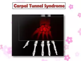
COMPRESSION OF MEDIAN NERVE IN CARPAL TUNNEL FLEXOR RETINACULUM Ulnar nerve does not pass through carpal tunnel MEDIAN NERVE CARPAL BONES SYNOVIAL SHEATH FLEXOR TENDON SWELLING OF SYNOVIAL SHEATHS PRODUCES COMPRESSION OF MEDIAN NERVE BECAUSE FLEXOR RETINACULUM AND CARPAL BONES ARE RIGID AND DO NOT STRETCH
SYMPTOMS OF CARPAL TUNNEL SYNDROME: MOTOR WEAKNESS OR PARALYSIS PARALYSIS OF MUSCLES OF THUMB - due to damage to Recurrent Branch of Median nerve Atrophy of muscles of thenar eminence - muscles at base thumb look flattened Loss of Opposition of Thumb OPPOSE
SYMPTOMS OF CARPAL TUNNEL SYNDROME: SENSORY LOSS Digital sensory branches of Median nerve SENSORY LOSS - anesthesia or numbness in distal part lateral palm; lateral 3.5 digits (thumb to lateral side of ring finger); on dorsal side, skin over the distal phalanges of same digits Note: Skin of proximal part of lateral palm may show no sensory loss (Palmar Cutaneous branch) Note: Palmar Cutaneous branch of Median Nerve does not pass through carpal tunnel
ULNAR NERVE IS NOT AFFECTED IN CARPAL TUNNEL SYNDROME ULNAR NERVES DOES NOT PASS THROUGH CARPAL TUNNEL Ulnar nerve innervates all other intrinsic muscles of hand (ex. interosseus muscles) Sensory innervation to medial palm and medial 1.5 digits ULNAR NERVE MEDIAN NERVE
RADIAL NERVE IS NOT AFFECTED IN CARPAL TUNNEL SYNDROME SENSORY MOTOR RADIAL NERVE - Innervates extensor muscles of arm and forearm (damage produces WRIST DROP) - Sensory innervation to dorsum of arm, forearm and hand In Hand: Radial nerve innervates no muscles - only sensory to dorsum of hand - does not pass through Carpal Tunnel RADIAL NERVE IN SPIRAL GROOVE RADIAL NERVE POSTERIOR ARM FOREARM: WRIST, FINGER EXTENSORS
ORDER OF STRUCTURES AT WRIST: LATERAL TO MEDIAL 4) Flexor digitorum superficialis and Palmaris longus tendons 1) Radial artery 2) Flexor carpi radialis tendon 5) Ulnar nerve and artery 3) Median nerve 6) Flexor carpi ulnaris tendon KNOW FOR SURGERY, REPAIR THUMB IS LATERAL