Acidos Nucleicos
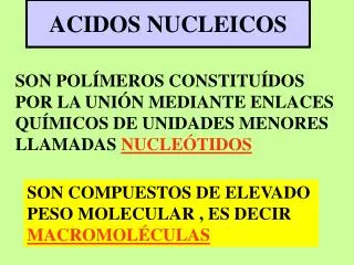
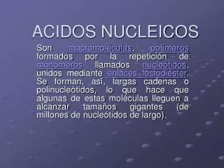
Acidos Nucleicos. Ácidos nucléicos. Ácidos nucléicos : un biopolímero que contiene tres tipos de monómeros unidos Una base orgánica heterociclo nitrogenado aromático derivado de purina y pirimidina Un azúcar monosacárido D-ribosa o 2-deoxi-D-ribosa ácido fosfórico

Acidos Nucleicos
E N D
Presentation Transcript
Ácidos nucléicos • Ácidos nucléicos : un biopolímero que contiene tres tipos de monómeros unidos • Una base orgánica heterociclo nitrogenado aromático derivado de purina y pirimidina • Un azúcar monosacárido D-ribosa o 2-deoxi-D-ribosa • ácido fosfórico • su nombre viene de que son muy abundantes en los núcleos celulares y de su carácter ácido • El más importente es el DNA que es el material genético. • En la siguiente diapositiva se muestra el nombre y abreviaturas de algunas bases nitrogenadas
NH 2 CH 3 NH 2 H N 2 Bases Purina/Pirimidina O O 4 3 HN HN N N 5 2 6 N N N N O O O 1 1 1 1 H H H Pirimidina Uracil (U) Timina (T) Citosina (C) O 7 6 N N 5 N 1 N HN N 8 N N N 2 N N N 4 9 3 H H H Purina Adenina (A) Guanina (G)
Nucleósidos • Nucleósido: son glucósidos cuya base son las nitrogenadas indicadas, contienen D-ribosa o 2-deoxy-D-ribose unidas a la bases por un enlace -N-glicosídico
O uracilo H N -D-ribosido 1 O Enlace-N glicosídico N 5' H O C H O 2 1' H H 4 ' H H carbono 2 ' 3 ' anomérico H O O H Uridina Nucleósidos • Uridina
Nucleótidos • Nucleótido: un nucleósido con una molecula de acido fofórico está esterificado con el -OH del monosacarido, más comúnmente el 3’-OH o el 5’-OH
Nucleótidos • Adenosina 5’-monofosfato (AMP)
Nucleótidos • Deoxitimidina 3’-monofosfato (3’dTMP)
H N C H 3 H N H N 2 H O C H H O C H 2 2 N 3 Aciclovir & AZT O O N N N O N O O H H H H H H H H H Azidotimidina (AZT) Aciclovir (Dibujado para ver su semejanza con la 2-deoxyguanosina
DNA - 1° Estructura • Ácidos Deoxirribonucleicos (DNA): un biopolíme-ro (polinucleótido) constituido por los ácidos desoxiadenílico, guanílico, citidílico y timidínico unidos por enlaces de éster fosfórico entre 3’-OH de una 2-deoxi-D-ribosa y el 5’-OH de la siguien-te 2-deoxi-D-ribosa. Son cadenas monótonas al-ternándose D-ribosa con fosfato y diferenciándo-se sólo en las bases colaterales a esa cadena. • Estructura Primaria :la secuencia de bases en el DNA determina su especificidad funcional y la información genética que almacena.
Extremo 5´fosforilado C H 3 H N - O - P - O - C H 2 - O H N H N 2 O = P C H 2 - O O H DNA – Estructura 1ia; TG O O O N 5' O O N H H 1' H H 2' O H N N 5' O O 1' H H H H Extremo 3´libre 3' 2' H
Estructura - 2° DNA • Estructura secundaria : es el ordenamiento de las cadenas de ácidos nucleicos • Hélice Doble : el DNA forma una doble hélice de dos cadenas complementarias con las vueltas a la derecha. Las bases están situadas hacia el in-terior de la hélice, en planos perpendiculares al eje, y los grupos de fosfórico están hacia el exte-rior. • Este modelo de doble hélice fue propuesto por James Watson y Francis Crick en 1953 en base a diagramas de rayos X y a las equivalencias A=T y G=C de las bases.
T-A Pareja de Bases • El mejor factor estabilizante es la pareja de bases T-A y entre C-G Timina Adenina
C-G Pareja de Bases Citosina Guanina
Formas del DNA • B-DNA • es la forma predominante en solución acuosa diluida • hélices con las vueltas a la derecha • 34 Å por 10 pares de bases; 20 Å de espesor • surco menor de 12 Å y el mayor de 22 Å • A-DNA • una helice diestra, pero de mayor espesor que B-DNA • 29 Å por 10 pares de bases
DNA – Estructura 3ia • EstructuraTerciaria : el arreglo de todos los átomos de ácido nucleico en la tercera dimensión, comúnmente referido como superenrollado • Circular DNA: un tipo de ADN double-stranded en el que 5 ' y 3 ' los finales de cada soporte son unidos según enlaces de fosfodiester (Fig 19.10) • Cromatina:consists de herida de moléculas de ADN alrededor de las partículas de histones en una estructura parecida a una cuenta
Ácidos Ribonucleicos (RNA) • RNA son similares al DNA en los que tambien son largas cadenas enroyadas de nucleotidos unidos por grupos de fosfodiester siendo el 3’-OH de una pentosa y el 5’-OH de la próxima. However, • the pentose unit in RNA is -D-ribose rather than -2-deoxy-D-ribose • the pyimidine bases in RNA are uracil and cytosine rather than thymine and cytosine • RNA is single stranded rather than double stranded
RNA • RNA molecules are classified according to their structure and function • Ribosomal RNA (rRNA): a ribonucleic acid found in ribosomes, the site of protein synthesis
RNA • Transfer RNA (tRNA): a ribonucleic acid that carries a specific amino acid to the site of protein synthesis on ribosomes
RNA • Messenger RNA (mRNA): a ribonucleic acid that carries coded genetic information from DNA to the ribosomes for the synthesis of proteins • present in cells in relatively small amounts and very short-lived • single stranded • its synthesis is directed by information encoded on DNA • a complementary strand of mRNA is synthesized along one strand of an unwound DNA, starting from the 3’ end
RNA • Transcription: the synthesis of mRNA from DNA
El Código Genético • Codon: a triplet of nucleotides on mRNA that directs incorporation of a specific amino acid into a polypeptide sequence
The Genetic Code • Properties of the Code • only 61 triplets code for amino acids; the remaining 3 (UAA, UAG, and UGA) signal chain termination • the code is degenerate, which means that several amino acids are coded for by more than one triplet. Leu, Ser, and Arg, for example, are each coded for by six triplets • for the 15 amino acids coded for by 2, 3, or 4 triplets, it is only the third letter of the codon that varies. Gly, for example, is coded for by GGA, GGG, GGC, and GGU • there is no ambiguity in the code; each triplet codes for one and only one amino acid
Sequencing DNA • Restriction endonuclease: an enzyme that catalyzes hydrolysis of a particular phosphodiester bond within a DNA strand • over 1000 endonucleases have been isolated and their specificities determined • typically they recognize a set sequence of nucleotides and cleave the DNA at or near that particular sequence • EcoRI from E. coli, for example, cleaves as shown
Sequencing DNA • examples of endonucleases
Sequencing DNA • Polyacrylamide gel electrophoresis: a technique so sensitive that it is possible to separate nucleic acid fragments differing from one another in only a single nucleotide • Chain termination or dideoxy method: a method developed by Frederick Sanger for sequencing DNA molecules
DNA Replication • the sequence of nucleotides on one strand is copied as a complementary strand to form the second strand of double-stranded DNA • this synthesis is catalyzed by the enzyme DNA polymerase • DNA polymerase will carry of this synthesis in vitro using single-stranded DNA as a template, provided the four dNTPs and a primer are present • because the new DNA strand grows from the 5’ to 3’ end, the primer must have a free 3’-OH group to which the first nucleotide of the growing chain is added
Chain Termin. Method • The key is addition of a 2’,3’-dideoxynucleoside triphosphate (ddNTP) to the synthesizing medium • synthesis terminates at any point where a ddNTP becomes incorporated
Chain Termin. Method • a single-stranded DNA of unknown sequence is mixed with primer and divided into four separate reaction mixtures • to each mixture is added all four dNTPs, one of which is labeled in its 5’- phosphoryl group with P-32 • also added are DNA polymerase and one of the four ddNTPs • when polyacrylamide gel electrophoresis of each reaction mixture is completed, a piece of x-ray film is placed over the gel to detect gamma radiation from the decay of P-32 • the base sequence of the complement to the original single-stranded template is read directly from the bottom to top of the developed film