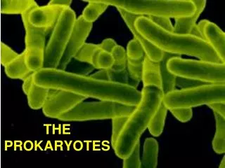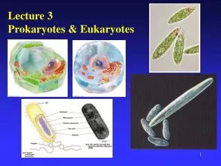
Chapter 3 The Prokaryotes
Chapter 3 The Prokaryotes. Chapter Outline. 3.1 Bacteria 3.2 Actinomycetes 3.3 Cyanobacteria 3.4 Archaeobacteria 3.5 Other prokaryotes 3.6 Classification of bacteria. Concepts.


Chapter 3 The Prokaryotes
E N D
Presentation Transcript
Chapter Outline 3.1 Bacteria 3.2 Actinomycetes 3.3 Cyanobacteria 3.4 Archaeobacteria 3.5 Other prokaryotes 3.6 Classification of bacteria
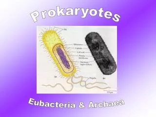
Concepts • Microorganisms are too small to be seen without the use of a microscope. The techniques-such as sterilization and the use of culture medium are required to isolate and grow these microbes. • Bacteria may be spherical (cocci), rod-shaped (bacilli), spiral, or filamentous. • Most bacteria can be divided into gram-positive and gram-negative groups based on their cell wall structure and response to the Gram stain. Bacteria such as mycoplasmas lack a cell wall.
3.1 Bacteria Size, Shape, and Arrangement of Bacterial Cells Most bacteria fall within a range from 0.2 to 2.0 μm in diameter and from 2 to 8μm in length. Cm = 10-2 meter mm = 10-3 meter μm = 10-6 meter nm = 10-9 meter They have a few basic shapes-spherical coccus (plural, cocci, meaning berries), rod-shaped bacillus (plural, bacilli, meaning little staffs), and spiral.
How to identify an unknown bacterial species ? • Morphology(shape) • Chemical composition(often detected by staining reactions) • Nutritional requirements • Biochemical activities • Source of energy(sunlight or chemicals) Factors:
The Micrococcaceae The family Micrococcaceae contains gram-positive cocci, 0.5-2.5 μm in diameter, that divide in more than one plane to form regular or irregular clusters of cells. All are aerobic or facultatively anaerobic. The peptidoglycan di-amino acid is L-lysine. The three most important genera are: • Micrococcus • Staphylococcus • Streptococcus
Micrococcus – aerobic, gram-positive, catalase positive, cell arranges mainly in pairs, tetrads, or irregular clusters, nonmotile. They are often yellow, orange or red in color
staphylococci staphylococci Staphylococcus - facultatively anaerobic, gram-positive, usually form irregular clusters, nonmotile, catalase positive but oxidase negative, ferment glucose anaerobically.
Streptococcus - facultatively anaerobic or microaerophilic, catalase negative, gram-positive, Cell arranges in pairs or chains, usually nonmotile, A few species are anaerobic rather than facultative.
Rod-shaped bacteria Bacilli divide only across their short axis, so there are fewer groupings of bacilli than of cocci. Single bacillus Diplobacilli streptobacilli Coccobacillus
Spore-forming rod shaped bacteria Almost all Spore-forming bacteria are Gram+ Bacillus – Aerobic Clostridium – Anaerobic Bacillus subtilis, B. Mycoides B. Pastturii B. megaterium B. Thuringiensis B. Anthracis B. Botulinus B. cereus Clostridium botulinus C. butyricum C. aceticum C. tetani C. putrificum
Nonspore - forming rod shaped bacteria Most nonspore – forming rod shaped bacteria are Gram - Representatives: Escherchia coli Alcaligenes Proteus Flavobacteria Pseudomonas Rhizobium Azotobacter
Vibrio, Spirillum and Spirochete Some bacteria are shaped like long rods twisted into spirals or helices; they are called vibrios (like commas or incomplete spirals), spirilla if rigid and spirochetes when flexable. vibrio spirillum spirochete
3.2 Actinomycetes Actinomycetes are filamentous bacteria. Their morphology resembles that of the filamentous fungi; however, the filaments of actinomycetes consist of procaryotic cells. Some actinomycetes resemble molds by forming externally carried asexual spores for reproduction. Filamentous, High G + C content, Gram-positive (63 – 78% GC)
Chain of conidiospores Aerial hyphae Agar surface Substrate mycelium The cross section of an actinomycete colony showing the substrate mycelium and aerial mycelium with chains of conidiospores
Various types of spore-bearing structures on the streptomyces
Antibiotics Actinomycetes Representive genera: Streptomyces Nocardia Actinomyces Micromonospora Streptosporangium Actinoplanes Frankia Over 500 distinct antibiotic substances have been shown to be produced by streptomycete. Most antibiotics are efficient against different bacteria. More than 50 antibiotics have been used in human and veterinary medicine, agriculture and industry
Chain of conidiospores Aerial hyphae Agar surface Substrate mycelium The cross section of an actinomycete colony showing the substrate mycelium and aerial mycelium with chains of conidiospores
Various types of spore-bearing structures on the streptomyces Streptomyces spores, called conidia, are not related in any way to the endospores of Bacillus and Clostridium because the streptomycete spores are produced simply by the formation of cross-walls in the multinucleate sporophores followed by separation of the individual cells directly into spores.
Ecology and isolation of Streptomyces: • Alkaline and neutral soils are more favorable for the development of Streptomyces than are acid soils. • Streptomyces require a lower water potential for growth than many other soil bacteria. • Media often selective for Streptomyces contain the usual assortment of inorganic salts
Concept • The streptonycetes are a large group of filamentous, gram positive bacteria that form spores at the end of aerial filaments. • They have the highest GC percentagein the DNA base composition of any bacteria known. • Many clinically important antibiotics have come from Streptomycetes species
3.3 Cyanobacteria The cyanobacteria have typical prokaryotic cell structures and a normal gram-negative cell wall. They range in diameter from about 1 – 10 µm and may be unicellular or form filaments. They have chlorophyll and carry out oxygen-producing photosynthesis, much as plants and the eukaryotic algae do.
Nonfilamentous cyanobacteria Filamentous Cyanobacterium,Anabaena sp. (SEM x5,000) The morphological diversity of the cyanobacteria is considerable. Both unicellular and filamentous forms are known, and considerable variation within these morphological types occurs.
Heterocysts have intercellular connections with adjacent vegetative cells, and there is mutual exchange of materials between these cells, with products of photosynthesis moving from vegetative cells to heterocysts and products of nitrogen fixation moving from heterocysts to vegetative cells.
Main function of Cyanobacteria • Photosynthesis • Nitrogen fixation • The cyanobacteria are the largest and most diverse group of photosynthetic bacteria. • The structure and physiology of the heterocyst ensures that it will remain anaerobic; it is dedicated to nitrogen fixation. It should be noted that nitrogen fixation also is carried out by cyanobacteria that lack heterocysts. • Cycnobacteria are capable of considerable metabolic flexibility.
Physiology of cyanobacteria: The nutrition of cyanobacteria is simple. Vitamins are not required, and nitrate or ammonia is used as nitrogen source. Nitrogen-fixing species are common. Most species tested are obligate phototrophs, However, some cyanobacteria are able to grow in the dark on organic compounds, using the organic material as both carbon and energy source.
Problems ! Many cyanobacteria produce potent neurotoxins, and during water blooms when massive accumulations of cyanobacteria may develop, animals ingesting such water may succumb rapidly.
3.4 The Archaebacteria Although archaebacteria are classified as procaryotes, these cells appear to be fundamentally different from typicaI bacteria or cyanobacteria. In fact, they represent a cell type that seems to be neither eucaryotic nor eubacterial.
The archaebacteria have the following unique combination of traits: Prokaryotic traits: • They are about 1 micrometer (um) in diameter, the size of typical procaryotes. • They lack membrane-bound organelles. • They have nuclear bodies (nucleoids) rather than true, menbranee bound nuclei. • Their ribosomes are 70 S, the size of those found in typical prokaryotes.
Eukaryotic traits: • Their cell walls completely lack peptidoglycan. • Their protein synthesis machinery is sensitive to inhibitors that typically affect only eukaryotes and is resistant to many inhibitors that affect prokaryotes. • Some of their proteins, pigments, and biochemical processes closely resemble those found in eukaryotic cells.
Archaebacteria include three groups: 1. The methanogens, strict anaerobes that produce methane (CH4) from carbon dioxide and hydrogen. 2. Extreme halophiles, which require high concentrations of salt for survival. 3. Thermoacidophiles, which normally grow in hot, acidic environments.
Methanogenic bacteria are strict anaerobes that obtain energy by converting C02, H2, formate, acetate, and other compounds to either methane or methane and C02. C02 + 4 H2 CH4 + 2H2O CH3 C00 H C02 + CH4
Sewage treatment plants use the methane produced to generate heat and electricity. Methanogenesis may eventually serve as a major source of pollution-free energy? !
Extremely thermophilic bacteria They are gram-negative, aerobic, irregularly lobed spherical bacteria with a temperature optimum around 70-80 0C and a pH optimum of 2 to 3. Their cell wall contains lipoprotein and carbohydrates but lacks peptidoglycan.
Extreme halophilic bacteria Their most distinctive characteristic is their requirement of a high concentration of sodium chloride for growth. They are aerobic chemoheterotrophs with respiratory metabolism and require complex nutrients, usually proteins and amino acids, for growth.
3.5 Other prokaryotes • Rickettsia • Chlamydia • Mycoplasma • Bdellovirio
Rickettsia 1. 0.2-0.5µm in diameter. obligate intracellular parasites. The majority of them are gram-negative and multiply only within host cells. 2. Binary fission within host cells.They lack the enzymatic capability to produce sufficient amounts of ATP to support their reproduction. They obtain the ATP from host cells. 3. Many species of them cause disease in humans and other animals.
Chlamydia • Obligate intracellular parasites, unable to generate sufficient ATP to support their reproduction. • Gram-negative and cell divides by binary fission • Cause humanrespiratory and genitourinary tract disease, and in birds they cause respiratory disease.
Mycoplasma • Diameter=0.1-0.25 µm. They lack cell wall, are bounded by a single triple-layered membrane. • They are the smallest organisms capable of self-reproduction. • The colony is “fried egg” appearance. • Several of them cause diseases in humans. (pneumonia, respiratory tract disease)
3.6 Classification of bacteria 1. MORPHOLOGICAL CHARACTERISTICS 2. DIFFERENTIAL STAINING 3. NUCLEIC ACID HYBRIDIZATION 4. NUMERICAL TAXONOMY
Fungi Plant Animal Protista Prokaryotae Five-kingdom system is a commonly accepted system of classification
Eukaryotes Archaebacteria Eubacteria Universal Phylogenetic Tree derived from comparative sequencing of 16S or 18S RNA. Note the threemajor domains of living organisms.
Divisions and Classes in the Kingdom Procaryotae (Monera) Identified by Common Names DIVISION CLASS Typical gram-negative cell wall Nonphotosynthetic bacteria Anaerobic photosynthetic bacteria Cyanobacteria Typical gram-positive cell wall Rods and cocci Actionmycetes and related organisms Mycopeanas Archaeobacteria Wall-less procaryotes Unusual walls
The taxonomic classification scheme for bacteria may be found in Bergey's Manual of Systematic Bacteriology. InBergey's Manual, bacteria are divided into four divisions. Three divisions consist of eubacterial cells, and the fourth division consists of the archaeobacteria. Each division is divided into classes
Classes are divided into orders families genera species Bacterial speciesis defined simply as a population of cells with similar characteristics. Strain is a group of cells all derived from a single cell.