Chapter 4 – Part B: Prokaryotic (bacterial) cells
260 likes | 450 Vues
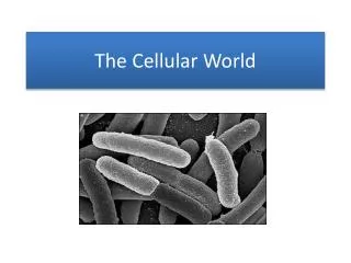
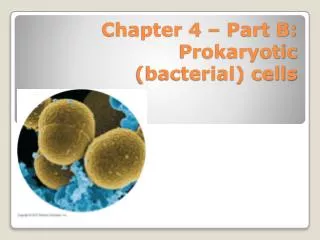
Chapter 4 – Part B: Prokaryotic (bacterial) cells. Prokaryote = bacteria Cells are smaller than eukaryotes, less organized, no membrane-bound organelles All molecules in these cells are in close contact with one another. General characteristics:. Parts:

Chapter 4 – Part B: Prokaryotic (bacterial) cells
E N D
Presentation Transcript
Prokaryote = bacteria • Cells are smaller than eukaryotes, less organized, no membrane-bound organelles • All molecules in these cells are in close contact with one another General characteristics:
Parts: 1. Cytoplasm --75% water for absorbing heat from chemical reactions; Dissolved and suspended molecules in the cytoplasm Cytoplasm
2. Nucleoid: Region where DNA is found (does not have a membrane around it!); DNA is 1 circular chromosome.
3. Plasmids: Extrachromosomal DNA Cell may have one copy or many Extra genes code for new traits, such as antibiotic resistance or production of toxin Passed to another cell by way of the sex pilus
plasmid Cell with plasmid and sex pilus Cell that will receive a copy of the plasmid 4. Sex pilus: 1 or 2 per cell containing plasmid(s) Long, hollow tube for transfer of a copy of a plasmid Transfer can happen between different species (highly unusual in nature) Made of protein called pilin
Cell with plasmid and sex pilus Cell with plasmid and sex pilus Conjugation
5. Ribosomes Site of protein synthesis 70S ribosomes in bacteria
6. Fimbriae Hair-like extensions from cell Also made of pilin, similar structure to sex pilus May have several or may cover the cell Important for attachment Ex. Neisseriagonorrhoeae infects urogenital tract by attaching to tissues there Image from: http://www.biosciednet.org/portal/search/searchResults.php?pageNumber=1&searchType=basic&sort=Relevance&pageNumber=1&searchType=basic&sort=Relevance&query=neisseria&gradeLevels=0
7. Flagellum Anchored in plasma membrane Made of flagellin protein (also called the H antigen) Ex. E. coli O157:H7 = strain of pathogenic E. coli Turns like a corkscrew (does not whip back and forth) Movement: Runs and tumbles More runs and fewer tumbles moving toward an attractant -taxis = movement chemotaxis = movement in response to chemical phototaxis = movement in response to light aerotaxis = in response to oxygen magnetotaxis = in response to Earth’s magnetic field Arrangement of flagella: monotrichous = 1 per cell amphitrichous = at both ends of cell lophotrichous = tuft at one end of cell peritrichous = covering cell Endoflagella: flagella wrapped around cell and covered with sheath Found in spirochetes
Salmonella movie Flagella
8. Cell envelope: A. Plasma membrane: Phospholipids and proteins Few molecules can move through hydrophobic phospholipids Many proteins regulate which molecules move into or out of the cell Function: Selectively permeable barrier Aerobic respiration Photosynthesis Enzymes for cell wall synthesis Attachment of chromosome during cell division Excretion/secretion Receptor sites (for recognition of molecules outside the cell) Cell envelope
B. Cell wall: Peptidoglycan …NAG – NAM – NAG – NAM… aa aa aa aa------aa aa aa aa …NAM – NAG – NAM – NAG… aa aa aa aa------aa aa aa aa …NAG – NAM – NAG – NAM… (NAG and NAM molecules are sugars; aa = amino acids) Cell wall
Lysozyme breaks the bonds between the NAG and NAM sugars Penicillin prevents the crossbridges between aa chains from forming Penicillin is only effective in actively growing cells Gram + cell wall can be 40 layers thick; G – is 1 or 2 layers thick Functions: strength, support, shape Cells without cell walls: • L-forms: bacteria that have lost their cell walls, many different species can do this • Mycoplasmas
C. Outer membrane (only in Gram – bacteria) Structure like the plasma membranes Contains proteins called porins Contains LPS – lipopolysaccharide Structure of LPS = Side chain is O Antigen Core Lipid A (buried in hydrophobic region of outer membrane); is an endotoxin You don’t want to lyse all Gram negative bacteria at once because of the danger of shock Function: an extra barrier Outer membrane of G- bacteria
D. Periplasmic space Space between membranes and cell wall Contains: Binding proteins:ex.To bind glucose molecules in environment Degrading enzymes: ex. To degrade macromolecules Detoxifying enzymes: ex. B-lactamase Periplasmic space
9. Endospores Protective structures; not reproductive structures Sporulation = 1 cell 1 spore Germination = 1 spore 1 cell Spore coat resistant to extreme environmental conditions: heat, dry, UV, chemicals, etc. Reason that we must use an autoclave to sterilize things Endospores
10. Capsule Also called glycocalyx, slime layer Mucus-like, sticky yet slippery Polysaccharide or polypeptide substance Function: Attachment, ex. Plaque on your teeth Movement – gliding Evasion of immune system Protection against dehydration Capsules
This is a photomicrograph of Streptococcus pneumoniae bacteria having been grown from a blood culture.Streptococcus pneumoniae, the bacteria responsible for pneumococcal meningitis, is very common, and normally lives in the back of the nose and throat, or the upper respiratory tract. http://phil.cdc.gov/phil/details.asp Photomicrograph of Streptococcus pneumoniae bacteria revealing capsular swelling using the Neufeld-Quellung test. This organism causes respiratory infections such as pneumonia and sinusitis, as well as bacteremia, otitis media, meningitis, peritonitis and arthritis. The Neufeld-Quellung test is used in pneumococcus typing. http://phil.cdc.gov/phil/details.asp