Download
1 / 13
130 likes | 351 Vues
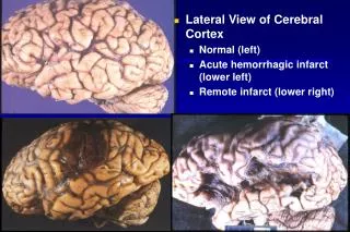
Lateral View of Cerebral Cortex Normal (left) Acute hemorrhagic infarct (lower left) Remote infarct (lower right). Coronal View of Cerebrum Normal (left) Acute hemorrhagic infarct (lower left) Remote infarct (lower right).

E N D
Lateral View of Cerebral Cortex • Normal (left) • Acute hemorrhagic infarct (lower left) • Remote infarct (lower right)
Coronal View of Cerebrum • Normal (left) • Acute hemorrhagic infarct (lower left) • Remote infarct (lower right)
Coronal section of cerebrum showing lacunar infarct in right internal capsule.
Brain showing superior sagittal sinus thrombosis and bilateral hemorrhagic infarcts.
Coronal section of cerebrum showing laminar necrosis of cortical ribbon.
Hypereosinophilic (red) neurons indicative of recent infarction and coagulative necrosis (HE stain).
Left: Vessels of the circle of Willis showing multiple aneurysms. Right: Inferior view of brain showing subarachnoid hemorrhage.
Coronal sections of cerebrum showing hemorrhage in thalamus (left) suggestive of hypertensive hemorrhage, and lobar hemorrhage (right) suggestive of congophilic amyloid angiopathy.
Coronal section of brain from a premature infant who died from germinal matrix hemorrhage with ventricular extension.
Coronal section of left occipital lobe (left) showing lobar hemorrhage and photomicrograph of leptomeningeal and parenchymal vessels showing birefringence following staining with congo red.
Subacute lateral medullary plate infarct and associated basilar artery showing atherosclerotic plaque with hemorrhage and occlusion of vessel.