The Hand
The Hand. Terminology. Anterior = Palmar (Volar also used) Posterior = Dorsal Ulnar (Medial) = pinky finger side Radial (Lateral) = thumb side. The Hand. Carpals. Phalanges. Metacarpals. Joints of Wrist & Hand. Radiocarpal joints Carpometacarpal joints Metacarpophalangeal joints

The Hand
E N D
Presentation Transcript
Terminology • Anterior = Palmar (Volar also used) • Posterior = Dorsal • Ulnar (Medial) = pinky finger side • Radial (Lateral) = thumb side
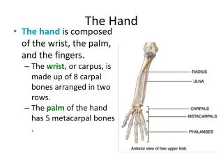
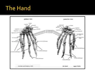
The Hand Carpals Phalanges Metacarpals
Joints of Wrist & Hand • Radiocarpal joints • Carpometacarpal joints • Metacarpophalangeal joints • Interphalangeal joints
Terminology • 27 bones per hand • 3 parts of hand • Proximal end, Carpals – Wrist • Hand proper, Metacarpals – Palm • Distal region, Phalanges – Fingers
Terminology • Anatomical position is PALM FORWARD • Humans retain ancestral (primitive) configuration of 5 sets of digits
Terminology • Wrist • 8 Carpals (carpus) form the wrist • 5 rays (I-V)—refer to metacarpals & phalanges (digits) • 5 metacarpals (metacarpus) • 14 phalanges • 5 proximal • 4 intermediate • Ray 1 does not have an intermediate phalanx (i.e., the thumb or pollex) • 5 distal
Terminology • Sesamoid bones (develop w/i tendons) • Usually found on palmar aspect of head for MC1
Ossification of Hand • Carpals • Single center of ossification • Metacarpals • 2 centers • Primary = shaft • Secondary = head • Except MC1 (secondary is at base) • Phalanges • 2 centers • Primary = shaft & distal end • Secondary = Base
The Carpus (Wrist) • 8 bones in 2 rows • very tightly bound by ligaments • Proximal Row (lateral – medial) • Scaphoid, lunate, triquetral, pisiform • First 2 articulate with the radius • Distal Row (lateral – medial) • Trapezium, trapezoid, capitate, hamate
The Carpus (Wrist) C H T T P T S L Palmar
Carpals—Scaphoid (Navicular of Hand) Hold like rubber ducky (or Donald Duck in profile) and the beak points to the side that it is from
Carpals—Lunate Place facet for the scaphoid on flat table with crescent moon facing you; the facet for the triquetral will “lean” to the side that it is from
Carpals—Triquetral (triquetrum) Place facet for the hamate (largest facet) on flat table with the facet for the pisiform and lunate facing you; the facet for the pisiform will be on the side that the bone is from
Carpals—Trapezium (Greater Multangular) Hold saddle facet superior with the trapezial ridge and groove facing you; the bone will “point” to the side that it is from
Carpals—Trapezoid (Lesser Multangular) Think of the traqpezoid as a boot for your foot. Visualize where the toes would go, the arch of your foot and the “zipper” on the lateral side. The side of your foot that the boot would “fit on is the side that it is from.
Carpals—Capitate Think of the capitate as Elvis’ head in profile. The facet for the hamate is his hair and the nonarticular palmar surface is his face. His hair is on the side that the bone is from.
Carpals—Hamate The hamate has a hook (hamulus). With the hook curved towards you and the facet for the capitate facing you, the hook will be positioned on the side that it is from.
Metacarpals • 5 bones numbered I-V, beginning laterally with the thumb • Round head, square-shaped base • Body (shaft) is slightly concave on the palmar side • Head is rounded for articulation with surface of proximal phalanges • Base has complex facets for multiple articulations with each other & with carpals
Metacarpal I • MC1- 1st metacarpal • short thick body, base articulates with trapezium only (single facet) • Saddle shaped sellar joint • Allows MCI to rotate 90° medially (for opposition of the thumb and fingers).
Metacarpal I L M Lateral facet = 0 Medial facet = 1 (very slight) Left Bone Depicted
Sesamoids • Small oval ossicles (extra bones) usually found where tendons traverse a joint between (2) bones • Most commonly on palmar surface (MC1 head)
Metacarpals II-V • Metacarpal II – usually longest, has wide & deeply cleft base • Metacarpal III – styloid process that projects proximally from the base • Metacarpal IV – Smaller than II and III, has a rectangular base • Metacarpal V – small, slight medial facet or tubercle
Metacarpal II Medial facet = 2 Lateral facet = 1 Left Bone Depicted
Metacarpal III Medial facet = 2 Lateral facet = 2 In anatomical position, styloid on side that the bone is from Left Bone Depicted
Metacarpal IV Medial facet = 1 Lateral facet = 2 Left Bone Depicted
Metacarpal V • Base of fifth metacarpal saddle shaped & articulates with hamate
Metacarpal V Medial facet = 0 Lateral facet = 1 Left Bone Depicted
Phalanges • 5 “rays” in 3 rows • Proximal • I-V • Intermediate • II-V • Distal • I-V
Proximal Phalanges • Body relatively long and smooth • Proximal end (base) concave • Distal end (head) convex
Intermediate Phalanges • Body relatively long and smooth • Shorter than proximal • Proximal end (base) concave • Double-faceted • Distal end (head) convex • Ray I (thumb/pollex) lacks intermediate phalanx
Distal Phalanges • Body short & flares out toward distal end • Base double-faceted • Distal (terminal)–diminutive flattened head, lacks distal articular facets • non-articular pad (distal phalangeal tuberosity)
Terminology • Plantar – sole of the foot • Dorsal – top of the foot • Proximal – toward the tibia • Distal – toward the toe
Feet • Foot bones are very similar to the hands, but slightly modified for: weight bearing, bipedal locomotion, shock absorbing and propulsion • 26 bones in each foot
Feet • 3 regions • Tarsals (Tarsus) – 7 bones, proximal portion of foot • Metatarsals (Metatarsus)– 5 bones, body of foot • Phalanges (toes)– 14 bones
Orientation of Tarsals & Metatarsals Right side depicted
Tarsals • Proximal row (2 bones) • Talus • Calcaneus • Intermediate row (1 bone) • Navicular • Distal row (4 bones) • Cuboid • Lateral cuneiform • Intermediate cuneiform • Medial cuneiform Right side depicted
Talus • Proximal row • Talus (astragalus in animals) • Articulates with distal tibia and fibula, calcaneus, and navicular. • Lacks muscle attachments Left side depicted
Talus Left side depicted
Talus Place on table with trochlea up and head away from you; it looks like a turtle! The foot is sticking out from the “shell” on the side that it is from.
Calcaneus (Heel) • Proximal row • Calcaneus – largest tarsal • Articulates with talus and cuboid. Left side depicted