Download

1 / 45
490 likes | 989 Vues
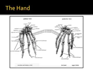
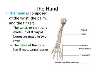
The Hand. The Palm of the Hand. Deep Fascia. The deep fascia of the wrist and palm is thickened to form the flexor retinaculum and the palmar aponeurosis . The palmar aponeurosis is triangular and occupies the central area of the palm

E N D
Deep Fascia • The deep fascia of the wrist and palm is thickened to form the flexor retinaculum and the palmaraponeurosis. • The palmaraponeurosis is triangular and occupies the central area of the palm • The apex of the palmaraponeurosis is attached to the distal border of the flexor retinaculum and receives the insertion of the palmarislongus tendon • The base of the aponeurosis divides at the bases of the fingers into four slips • Each slip divides into two bands, one passing superficially to the skin and the other passing deeply to the root of the finger • each deep band divides into two, which diverge around the flexor tendons and finally fuse with the fibrous flexor sheath and the deep transverse ligaments. • The medial and lateral borders of the palmaraponeurosis are continuous with the thinner deep fascia covering the hypothenar and thenar muscles • From each of these borders, fibrous septa pass posteriorly into the palm and take part in the formation of the palmarfascial spaces • The function of the palmaraponeurosis is to give firm attachment to the overlying skin and so improve the grip and to protect the underlying tendons.
The Carpal Tunnel • The carpus is deeply concave on its anterior surface and forms a bony gutter. The gutter is converted into a tunnel by the flexor retinaculum • The long flexor tendons to the fingers and thumb pass through the tunnel and are accompanied by the median nerve • The four separate tendons of the flexor digitorumsuperficialis muscle are arranged in anterior and posterior rows, those to the middle and ring fingers lying in front of those to the index and little fingers • At the lower border of the flexor retinaculum, the four tendons diverge and become arranged on the same plane • The tendons of the flexor digitorumprofundus muscle are on the same plane and lie behind the superficialis tendons. • All eight tendons of the flexor digitorumsuperficialis and profundusinvaginate a common synovial sheath from the lateral side • The tendon of the flexor pollicislongus muscle runs through the lateral part of the tunnel in its own synovial sheath • The median nerve passes beneath the flexor retinaculum in a restricted space between the flexor digitorumsuperficialis and the flexor carpiradialis muscles
Anatomical snuffbox • The 'anatomical snuffbox' is a term given to the triangular depression formed on the posterolateral side of the wrist and metacarpal I by the extensor tendons passing into the thumb • The base of the triangle is at the wrist and the apex is directed into the thumb. The impression is most apparent when the thumb is extended: • the lateral border is formed by the tendons of the abductor pollicislongus and extensor pollicisbrevis • the medial border is formed by the tendon of the extensor pollicislongus; • the floor of the impression is formed by the scaphoid and trapezium, and distal ends of the tendons of the extensor carpiradialislongus and extensor carpiradialisbrevis. • The radial artery passes obliquely through the anatomical snuffbox, deep to the extensor tendons of the thumb and lies adjacent to the scaphoid and trapezium. • Terminal parts of the superficial branch of the radial nerve pass subcutaneously over the snuffbox as does the origin of the cephalic vein from the dorsal venous arch of the hand.
Fibrous Flexor Sheaths • The anterior surface of each finger, from the head of the metacarpal to the base of the distal phalanx, is provided with a strong fibrous sheath that is attached to the sides of the phalanges • The proximal end of the fibrous sheath is open, whereas the distal end of the sheath is closed and is attached to the base of the distal phalanx • The sheath and the bones form a blind tunnel in which the flexor tendons of the finger lie. • In the thumb, the osteofibrous tunnel contains the tendon of the flexor pollicislongus • In the case of the four medial fingers, the tunnel is occupied by the tendons of the flexor digitorumsuperficialis and profundus • The fibrous sheath is thick over the phalanges but thin and lax over the joints.
Synovial Flexor Sheaths • the tendons of the flexor digitorumsuperficialis and profundus muscles invaginate a common synovial sheath from the lateral side • The medial part of this common sheath extends distally without interruption on the tendons of the little finger • The lateral part of the sheath stops abruptly on the middle of the palm, and the distal ends of the long flexor tendons of the index, the middle, and the ring fingers acquire digital synovial sheaths as they enter the fingers • The flexor pollicislongus tendon has its own synovial sheath that passes into the thumb • These sheaths allow the long tendons to move smoothly, with a minimum of friction, beneath the flexor retinaculum and the fibrous flexor sheaths. • The synovial sheath of the flexor pollicislongus (sometimes referred to as the radial bursa) communicates with the common synovial sheath of the superficialis and profundus tendons (sometimes referred to as the ulnar bursa) at the level of the wrist in about 50% of subjects • The vinculalonga and brevia are small vascular folds of synovial membrane that connect the tendons to the anterior surface of the phalanges and convey blood vessels to the tendons.
Insertion of the Long Flexor Tendons • Each tendon of the flexor digitorumsuperficialis enters the fibrous flexor sheath • opposite the proximal phalanx it divides into two halves, which pass around the profundus tendon and meet on its deep or posterior surface, where partial decussation of the fibers takes place • The superficialis tendon, having united again, divides almost at once into two further slips, which are attached to the borders of the middle phalanx. • Each tendon of the flexor digitorumprofundus, having passed through the division of the superficialis tendon, continues downward, to be inserted into the anterior surface of the base of the distal phalanx
Small Muscles of the Hand • The small muscles of the hand include the • four lumbrical muscles, • the eight interossei muscles, • the short muscles of the thumb, • and the short muscles of the little finger
Key • MCP= metacarpophalangial joints • CMC= carbametacarpal joints • PIP= proximal inter-phalangial joint • DIP= Distal interphalangial joint • ABD= Abduction • ADD= Adduction
Palmar Interossei • O • 1st – ulnar side base of 1st metacarpal bone • 2nd – ulnar side of 2nd MC bone • 3rd – radial side of 4th MC bone • 4th – radia side of 5th MC bone • I • Extensor expansion of 2,4 and 5th digits • N • Ulnar • F • ADD of 1st, 2nd, 4th and 5th digits toward midline of hand
Dorsal Interossei • O • 1st lateral head – ulnar side of 1st metacarpal bone • 1st medial head – radial side of 2nd metacarpal bone • 2nd, 3rd, 4th space between metacarpal bones • I • 1st – radial side 2nd proximal phalanx • 2nd – radial side of 3rd • 3rd – ilnar side of 3rd • 4th – ulnar side of 4th • N • Ulnar • F • ABD of 2nd, 3rd, and 5th finger from midline
Lumbricales • O • Tendons of FDP • I • Extensor expansion on dorsal aspect of each digits radial side • N • 1 and 2 – median • 3 and 4 – ulnar • F • MCP flexion 2-5 digits • DIP & PIP ext 2-5 digits
Palmaris Brevis • O • Flexor retinaculum • I • Palmar surface skin on ulnar side of hand • N • Ulnar • F • Wrinkles skin of hand on ulnar side
The short muscles of the thumb are the abductor pollicis brevis, the flexor pollicis brevis, the opponens pollicis, and the adductor pollicis The first three of these muscles form the thenar eminence. Thenar Eminance
Adductor Pollicis • O • Oblique Head • Capitate bone • Bases of 2-3 metacarpals • Transverse Head • Proximal 2/3 of palmar surface of 3rd metacarpal • I • Ulnar side of base of 1st proximal phalanx • N • Ulnar • F • CMC ADD of thumb
Abductor Pollicis Brevis • O • Scaphoid tuberosity • Trapezium ridge • Transverse carpal ligament • I • Lateral base f proximal 1st phalanx • N • Median • F • CMC & MCP ABD of thumb
Flexor Pollicis Brevis • O • Superficial head – trapezium • Deep head – trapezoid, capitate and palmar ligaments of distal carpal bones • I • Base of prximal 1st phalanx on radial side • Extensor expansion • N • Superficial – median • Deep – Ulnar • F • CMC & MCP Flexion of thumb
Opponens Pollicis • O • Trapezium • Transverse Carpal Ligament • I • Radial side of 1st metacarpal shaft • N • Median • F • Opposition
Opposition of the Thumb • the opponenspollicis muscle pulls the thumb medially and forward across the palm • so that the palmar surface of the tip of the thumb may come into contact with the palmar surface of the tips of the other fingers • It is an important muscle and enables the thumb to form one claw in the pincerlike action used for picking up objects • This complex movement involves a flexion of the carpometacarpal and metacarpophalangeal joints and a small amount of abduction and medial rotation of the metacarpal bone at the carpometacarpal joint
Abduction and Adduction of the Thumb • Abduction of the thumb may be defined as a movement forward of the thumb in the anteroposterior plane • It takes place at the carpometacarpal joint and the metacarpophalangeal joint. • Adduction can be defined as a movement backward of the abducted thumb in the anteroposterior plane. • It restores the thumb to its anatomic position, which is flush with the palm. • The adductor pollicis is the muscle that, in association with the flexor pollicislongus and the opponenspollicis muscles, is largely responsible for the power of the pincers grip of the thumb. • Adduction of the thumb occurs at the carpometacarpal and at the metacarpophalangeal joint.
Short Muscles of the Little Finger • The short muscles of the little finger are the abductor digiti minimi, the flexor digiti minimi brevis, and the opponens digiti minimi, which together form the hypothenar eminence • The opponens digiti minimi muscle is only capable of rotating the fifth metacarpal bone to a slight degree • However, it assists the flexor digiti minimi in flexing the carpometacarpal joint of the little finger • thereby pulling the fifth metacarpal bone forward and cupping the palm.
Intrinsic Hand Muscles Hypothenar Eminence
Abductor Digiti Minimi • O • Pisiform • I • Ulnar side base of 5th proximal phalanx • N • Ulnar • F • MCP ABD of 5th digit
Opponen Digiti Minimi • O • Hook of hamate • Transverse carpal ligament • I • Ulnar border of entire 5th metacarpal bone • N • Ulnar • F • MCP flexion & rotation of 5th digit
Flexor Digiti Minimi • O • Hamate bone • Transverse carpal ligament • I • Ulnar side of proximal 5th phalanx • N • Ulnar • F • MCP Flexion of 5th digit
Fascial Spaces of the Palm • The thenar space contains the first lumbrical muscle and lies posterior to the long flexor tendons to the index finger and in front of the adductor pollicis muscle • The midpalmar space contains the second, third, and fourth lumbrical muscles and lies posterior to the long flexor tendons to the middle, ring, and little fingers • The lumbrical canal is a potential space surrounding the tendon of each lumbrical muscle and is normally filled with connective tissue
Arteries of the Palm • Ulnar Artery • The ulnar artery enters the hand anterior to the flexor retinaculum on the lateral side of the ulnar nerve and the pisiform bone • The artery gives off a deep branchand then continues into the palm as the superficial palmar arch. • The superficial palmar arch is a direct continuation of the ulnar artery • On entering the palm, it curves laterally behind the palmaraponeurosis and in front of the long flexor tendons • The arch is completed on the lateral side by one of the branches of the radial artery • The curve of the arch lies across the palm, level with the distal border of the fully extended thumb • The deep branch of the ulnar artery arises in front of the flexor retinaculum, passes between the abductor digitiminimi and the flexor digitiminimi • joins the radial artery to complete the deep palmar arch
Radial Artery • On entering the palm, it curves medially between the oblique and transverse heads of the adductor pollicis and continues as the deep palmar arch • The deep palmar arch is a direct continuation of the radial artery • It curves medially beneath the long flexor tendons and in front of the metacarpal bones and the interosseous muscles • The arch is completed on the medial side by the deep branch of the ulnar artery • The curve of the arch lies at a level with the proximal border of the extended thumb. • The deep palmar arch sends branches superiorly, which take part in the anastomosis around the wrist joint, and inferiorly, to join the digital branches of the superficial palmar arch.
Branches of the Radial Artery in the Palm • Immediately on entering the palm, the radial artery gives off the arteria radialis indicis, which supplies the lateral side of the index finger • and the arteria princeps pollicis, which divides into two and supplies the lateral and medial sides of the thumb.
Allen's test • To test for adequate anastomoses between the radial and ulnar arteries, compress both the radial and ulnar arteries at the wrist, then release pressure from one or the other, and determine the filling pattern of the hand • If there is little connection between the deep and superficial palmar arteries only the thumb and lateral side of the index finger will fill with blood (become red) when pressure on the radial artery alone is released.
Veins of the Palm • Superficial and deep palmar arterial arches are accompanied by superficial and deep palmar venous arches, receiving corresponding tributaries.
Lymph Drainage of the Palm • The lymph vessels of the fingers pass along their borders to reach the webs • From here the vessels ascend onto the dorsum of the hand • The lymph from the medial side of the hand ascends in vessels that accompany the basilic vein • they drain into the supratrochlear nodes and then ascend to drain into the lateral axillary nodes. • The lymph from the lateral side of the hand ascends in vessels that accompany the cephalic vein • they drain into the infraclavicular nodes, and some drain into the lateral axillary nodes.
Nerves of the Palm • Median Nerve • The median nerve enters the palm by passing behind the flexor retinaculum and through the carpal tunnel. • It immediately divides into lateral and medial branches • The muscular branch takes a recurrent course around the lower border of the flexor retinaculum and lies about one fingerbreadth distal to the tubercle of the scaphoid • it supplies the muscles of the thenar eminence (the abductor pollicisbrevis, the flexor pollicisbrevis, and the opponenspollicis) and the first lumbrical muscle. • The cutaneous branches supply the palmar aspect of the lateral three and a half fingers and the distal half of the dorsal aspect of each finger • One of these branches also supplies the second lumbrical muscle. • the palmarcutaneous branch of the median nerve given off in the front of the forearm • crosses anterior to the flexor retinaculum and supplies the skin over the lateral part of the palm
Ulnar Nerve • The ulnar nerve enters the palm anterior to the flexor retinaculum alongside the lateral border of the pisiform bone • As it crosses the retinaculum it divides into a superficial and a deep terminal branch. • The superficial branch of the ulnar nerve descends into the palm, lying in the subcutaneous tissue between the pisiform bone and the hook of the hamate • The ulnar artery is on its lateral side. Here, the nerve and artery may lie in a fibro-osseous tunnel, the tunnel of Guyon, created by fibrous tissue derived from the superficial part of the flexor retinaculum • The nerve may be compressed at this site, giving rise to clinical signs and symptoms. • The nerve gives off the following branches: • a muscular branch to the palmarisbrevis • cutaneous branches to the palmar aspect of the medial side of the little finger and the adjacent sides of the little and ring fingers • It also supplies the distal half of the dorsal aspect of each finger.
Deep Branch of the Ulnar Nerve • The deep branch of the ulnar nerve runs backward between the abductor digiti minimi and the flexor digiti minimi • It pierces the opponens digiti minimi, winds around the lower border of the hook of the hamate, and passes laterally within the concavity of the deep palmar arch. • The nerve lies behind the long flexor tendons and in front of the metacarpal bones and interosseous muscles • It gives off muscular branches to the three muscles of the hypothenar eminence • the abductor digiti minimi, the flexor digiti minimi, and the opponens digiti minimi. • It supplies all the palmar and dorsal interossei, the third and fourth lumbrical muscles, and both heads of the adductor pollicis muscle.
The palmar cutaneous branch of the ulnar nerve given off in the front of the forearm crosses anterior to the flexor retinaculum • and supplies the skin over the medial part of the palm
The Dorsum of the Hand • The skin on the dorsum of the hand is thin, hairy, and freely mobile on the underlying tendons and bones. • The sensory nerve supply to the skin on the dorsum of the hand is derived from the superficial branch of the radial nerve and the posterior cutaneous branch of the ulnar nerve. • The superficial branch of the radial nerve winds around the radius deep to the brachioradialis tendon, descends over the extensor retinaculum, and supplies the lateral two thirds of the dorsum of the hand • divides into several dorsal digital nerves that supply the thumb, the index and middle fingers, and the lateral side of the ring finger • The area of skin on the back of the hand and fingers supplied by the radial nerve is subject to variation • Frequently, a dorsal digital nerve, a branch of the ulnar nerve, also supplies the lateral side of the ring finger
The posterior cutaneous branch of the ulnar nerve winds around the ulna deep to the flexor carpi ulnaris tendon • descends over the extensor retinaculum, and supplies the medial third of the dorsum of the hand • It divides into several dorsal digital nerves that supply the medial side of the ring and the sides of the little fingers • The dorsal digital branches of the radial and ulnar nerves do not extend far beyond the proximal phalanx • The remainder of the dorsum of each finger receives its nerve supply from palmar digital nerves.
Dorsal Venous Arch • The dorsal venous arch lies in the subcutaneous tissue proximal to the metacarpophalangeal joints • drains on the lateral side into the cephalic vein and, on the medial side, into the basilic vein • the greater part of the blood from the whole hand drains into the arch, which receives digital veins • freely communicates with the deep veins of the palm through the interosseous spaces.
Insertion of the Long Extensor Tendons • The four tendons of the extensor digitorum emerge from under the extensor retinaculum and fan out over the dorsum of the hand • The tendons are embedded in the deep fascia, and together they form the roof of a subfascial space, which occupies the whole width of the dorsum of the hand • Strong oblique fibrous bands connect the tendons to the little, ring, and middle fingers, proximal to the heads of the metacarpal bones • The tendon to the index finger is joined on its medial side by the tendon of the extensor indicis • the tendon to the little finger is joined on its medial side by the two tendons of the extensor digitiminimi
On the posterior surface of each finger, the extensor tendon joins the fascial expansion called the extensor expansion • Near the proximal interphalangeal joint, the extensor expansion splits into three parts: a central part, which is inserted into the base of the middle phalanx, • two lateral parts, which converge to be inserted into the base of the distal phalanx • The dorsal extensor expansion receives the tendon of insertion of the corresponding interosseous muscle on each side • distally receives the tendon of the lumbrical muscle on the lateral side
The Radial Artery on the Dorsum of the Hand • The radial artery winds around the lateral margin of the wrist joint, beneath the tendons of the abductor pollicis longus and extensor pollicis brevis, and lies on the lateral ligament of the joint • On reaching the dorsum of the hand, the artery descends beneath the tendon of the extensor pollicis longus to reach the interval between the two heads of the first dorsal interosseous muscle • the artery turns forward to enter the palm of the hand • Branches of the radial artery on the dorsum of the hand take part in the anastomosis around the wrist joint. Dorsal digital arteries pass to the thumb and index finger