Effects of Kinase Inhibitors on KHT Cell Scattering: Supplemental Figure 1
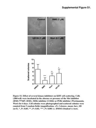
Supplemental Figure 1 illustrates the impact of various kinase inhibitors—BMS-777607 (Met inhibitor), U0126 (MEK inhibitor), and Wortmannin (PI3K inhibitor)—on KHT cell scattering. Cells were incubated for 4 days with or without the inhibitors at specified concentrations. Photographs of cell colonies were taken, and scattered colonies were counted from five random fields (magnification x5). Results are expressed as mean ± standard deviation (n=5), with statistical significance indicated (P < 0.05, P < 0.01, P < 0.001 vs DMSO, using Student's t-test).

Effects of Kinase Inhibitors on KHT Cell Scattering: Supplemental Figure 1
E N D
Presentation Transcript
BMS (1 M) Control U0126 (1 M) Wort (0.1 M) Supplemental Figure S1. Figure S1. Effect of several kinase inhibitors on KHT cell scattering. Cells (200/well) were incubated in the absence or presence of the Met inhibitor (BMS-777607, BMS), MEK inhibitor (U1026) or PI3K inhibitor (Wortmannin, Wort) for 4 days. Cell colonies were photographed and scattered colonies were counted from 5 random fields (magnification, 5). Columns, mean; bars, SD (n=5). *, P< 0.05; **, P< 0.01; ***, P< 0.001 vs. DMSO (Student’s t-test).