Burn Injuries
Burn Injuries. Chantel R. Grubbs Robert Smith Latrice Wilson Gabrielle Paul . Case Presentation PHA 5601 Pediatrics Ambulatory Care Dr. Angela Thornton, PharmD March 21 st , 2013. Objectives. Classify the types of burns, along with defining the prevalence, causes, and pathophysiology.

Burn Injuries
E N D
Presentation Transcript
Burn Injuries Chantel R. Grubbs Robert Smith Latrice Wilson Gabrielle Paul Case Presentation PHA 5601 Pediatrics Ambulatory Care Dr. Angela Thornton, PharmD March 21st, 2013
Objectives Classify the types of burns, along with defining the prevalence, causes, and pathophysiology. Explain various preventative measures, and different disabilities that may arise as a result of burn. Outline the approach to management and treatment options. Discuss and compare related clinical trials on various medication used to treat pediatric burns.
Definition A burn is an injury caused by fire, heat, radiation, electricity, or flammable agent.
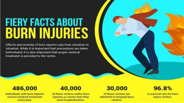
Epidemiology It is estimated that 1.2 million people in the United States require medical care for burn injuries each year, with 51,000 requiring hospitalization. About 30-40% of those patients are less than 15 years old. With the average age of the patient being only 2.5 years of age. The most common burn injuries result from exposure to heat and chemicals. It is the second leading cause of death for children under the age of 12.
Risk Factors Child negligence Child abuse Improper Adult supervision Cognitive Impairment Psychiatric illness
Pathophysiology of Thermal Burns Thermal Burn Injury Pathophysiology is based off of Jackson’s Thermal Wound Theory. The zone of coagulation is the area near the burn cell membranes rupture, clotted blood and thrombosed vessels. Zone of Stasis area surrounds the zone of coagulation inflammation, and is characterized by decreased blood flow. Zone of Hyperemia is the peripheral area of burn characterized by increased blood flow.
Pathophysiology of Electrical Burns • There are 3 proposed mechanisms of electrical burns: • Electrical energy causing direct tissue damage, altering cell membrane resting potential. • Conversion of electrical energy into thermal energy, causing massive tissue destruction and coagulation necrosis. • Mechanical injury with direct trauma resulting from falls or violent muscle contraction.
Pathophysiology of Chemical Burns • There are 6 proposed mechanisms by which chemicals cause burn-like injuries: • Oxidation: The protein denaturation is caused by inserting an oxygen, sulphur, or halogen atom to viable body proteins. • Reduction: Reducing agents act by binding free electrons in tissue proteins. Heat may also be a product of a chemical reaction, thereby causing a mixed picture • Corrosion: It causes protein denaturation on contact. They tend to produce a soft, which may progress to shallow ulceration
Pathophysiology of Chemical Burns • Mechanisms Continued: • Protoplasmic poisons: They produce their effects by causing the formation of esters with proteins or by binding or inhibiting calcium or other organic ions necessary for tissue viability and function. • Vesicants: They produce ischemia with anoxic necrosis at the site of contact. These agents are characterized to produce cutaneous blisters. • Desiccants: These substances cause damage by dehydration of tissues. The damage is often exacerbated by heat production, as these reactions are usually exothermic.
Etiology Chemical Burns Radiation Burns Electrical Burns Thermal Burns
Types of Burns 13 • First-degree(superficial burn)burns affect only the outer layer, also known as the epidermis. • Second-degree (partial thickness) burns affect both the epidermis and a variable portion of the dermal layer. • Superficial partial • Mid partial
Types of Burns 14 • Third-degree (full thickness) burns affect the entire epidermis, dermis and subcutaneous layers. • Deep partial • Full thickness • Fourth-degree (full thickness) burns affect the epidermis, dermis, muscle, tendon, and bone.
Clinical Manifestations • First Degree • Dry • No blisters • Edema • Erythema
Clinical Manifestations • Second Degree • Moist • Blisters • Underlying tissue is marked with pink and white spots • Fair capillary refill • Bleeds 17
Clinical Manifestations Superficial Partial Mid Partial
Clinical Manifestations • Third Degree • No bleeding • Dry • Presents with white, brown and black markings • Waxy 19
Clinical Manifestations Deep Partial Thickness Full Thickness
Clinical Manifestations • Fourth Degree • Muscles and bone is visible • Blackened and charred appearance 21
Differential Diagnosis • Steven’s Johnson’s Syndrome • Anticonvulsants • Carbemazepine (Tegretol®) • Valproate (Depakote®) • Zonisamide (Zonegran®) • Sulfonamides • Bactrim® • Antibiotics • Penicillins • Toxic Epidermal Necrolysis
Diagnosis • Burn severity is dictated by Percent total body surface area (TBSA) involvement • Rule of Nines • Rule of Palm 24
Dispelling Myths About Burn Treatment Toothpaste Ice Butter Egg Whites Bursting Blisters
Goals of Therapy Prevention of infection Acute care and resuscitation Wound management Pain relief Reconstruction Rehabilitation and Psychosocial adjustment
First Aid Treatment • Extinguish the Fire • Bring child to more ventilated area • Carefully remove smoldering clothing and jewelry • Thoroughly irrigate area with cool water to remove an access particles or debris • Cover area with a clean dry sheeting and apply cold wet compress to small injuries • Administer OTC analgesic • Acetaminophen (Tylenol®) • 10-15 mg/kg every 4-6 hours • Ibuprofen (Advil®) • 5-10 mg/kg every 6-8 hours
Nonpharmacological Aloe Vera Cool compresses
Aspects of Treatment Topical Agents Pain Management Antihistamines Fluid Resuscitation Nutritional Support Wound Closures Respiratory Therapy
Treatment of Superficial 1st Degree & Superficial Dermal 2nd Degree Burns
Treatment of Superficial 1st Degree & Superficial Dermal 2nd Degree Burns • First and second degree burns on <10% of the body may be treated with outpatient therapy • There is an increase risk of tetanus following a burn injury • Be sure to administer vaccine if patient has not been already vaccinated.
Treatment of Superficial 1st Degree & Superficial Dermal 2nd Degree Burns • Blisters should be left in tact and not be popped and treated with topical agents • Bacitracin (AK-Tracin®) ($8.00) -Prescription • Silver Sulfadiazine Cream (Silvadene®) ($20.00)-Prescription • Dressings should be changed daily • Wound should we washed with lukewarm water. • Small 1st degree and mild 2nd degree burn on face can be treated with Bacitracin and left open • Vaseline($4.00)- OTC
Treatment of Superficial 1st Degree & Superficial Dermal 2nd Degree Burns Silver Sulfadiazine
Proper Dressing Application Wash hands Carefully remove old dressing and throw away Clean burn area mild soap (ie. Hibiclens) and lukewarm or cool water Rinse area thoroughly Pat dry with clean towel or gauze Check for healing Apply prescribed ointment or fresh dressing Wrap sterile gauze over dressing to secure
Dressings for Ruptured Blisters • Ruptured blisters are treated with wound dressing/wound membrane • AQUACEL- Ag • Biobrane • Mepilex Ag • Acticoat • Hydrocolloid dressings • Impregnated gauze • Dressing are kept on for 7-10 days and check periodically through the week • All dressings that are on the formulary are found in the burn unit supply closet.
Dressings • Hydrocolloid Dressing • DuoDERM • Tegaderm • Comfeel • Nu-Derm • Impregnated Gauze • Xeroform impregnated gauze • Chlorhexidine impregnated gauze (Bactigras) • Petrolatem/Vaseline impregnated gauze
Dressings Hydrocolloid Dressings
Dressings Impregnated gauze
Topical Agents • Topical agents for more severe burns • AQUACEL Ag • Available by Prescription • Sulfacetamide Acetate* • Silver Sulfadiazine • Mafenide Acetate - • 0.5% Silver Nitrate Solution • Accuzyme Ointment *
Criteria for Hospital Admission Burns affecting >15% of body surface area Electrical burns caused by high-tension wires Inhalation injury, regardless of the amount of body surface area burned Inadequate home situation Suspected child abuse or neglect Burns to the hands, feet, or genitals
Fluid Resuscitation • Lactated Ringer Solution • Dose • Parkerland Formula: 4 mL lactated Ringer/kg/% BSA burned • Half of fluid given 1st 8hrs; Next half administered over 16hrs • IV Infusion Rate Varies • Monitoring Parameters • Urine Output • Glucose • Clinical Pearls • Initial fluid should be warmed • Infants are at risk for hypoglycemia
Fluid Resuscitation • Albumin (Albuked 25 ®, Flexbumin 25%®, Human Albumin Grifols 25%®, Kedbumin®, Plasbumin2-25®, Plasbumin-5®) • Use 5% albumin solution • 30–50% of total BSA, 0.3 mL /kg/% BSA • 50–70% of total BSA, 0.4 mL/kg/% BSA • 70–100% of total BSA, 0.5 mL/kg/% BSA burn • Monitoring Parameter: Albumin≥2g/dl • Clinical Pearls • Concurrent use for burn >85% of BSA • Administered 8-24hr after injury
Qualifications for Albumin Treatment • It is not usually screened for infections. Albumin is synthesized exclusively in the liver. The normal rate of albumin synthesis is 0.2g/kg body weight per day. • Controversy exists as to whether colloid should be provided in the early period of burn resuscitation. One preference is to use colloid replacement concurrently if the burn is >85% of total BSA. Colloid treatment is usually instituted 8–24 hr after the burn injury. • For burns of 30–50% of total BSA, 0.3 mL of 5% albumin/kg/% BSA burn is infused over a 24-hr period • For burns of 50–70% of total BSA, 0.4 mL/kg/% BSA burn is infused over 24 hr • For burns of 70–100% of total BSA, 0.5 mL/kg/% BSA burn is infused over 24 hr