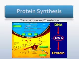
Protein synthesis
Protein synthesis. 30.4 Ribosome Structure and Assembly 30.5 Mechanics of Protein Synthesis. 30.4 Ribosome Structure and Assembly. E. coli ribosome is 25 nm diameter, 2520 kD in mass, and consists of two unequal subunits that dissociate at < 1mM Mg 2+

Protein synthesis
E N D
Presentation Transcript
Protein synthesis • 30.4 Ribosome Structure and Assembly • 30.5 Mechanics of Protein Synthesis
30.4 Ribosome Structure and Assembly • E. coli ribosome is 25 nm diameter, 2520 kD in mass, and consists of two unequal subunits that dissociate at < 1mM Mg2+ • 30S subunit is 930 kD with 21 proteins and a 16S rRNA • 50Ssubunit is 1590 kD with 31 proteins and two rRNAs: 23S rRNA and 5S rRNA • These ribosomes and others are roughly 2/3 RNA • 20,000ribosomes in a cell, 20% of cell's mass
Ribosomal RNA • 3 rRNA molecules • 23S, 16S, 5S • Derived from a single 30S rRNA precursor transcript • Extensive intrachain H-bonding • 2/3 rRNA is helical
Ribosomal Proteins • One of each per ribosome, except L7/L12 with 4 • L7/L12 identical except for extent of acetylation at N-terminus • Only one protein is common to large and small subunits: S20 = L26 • Variety of structures, still being characterized
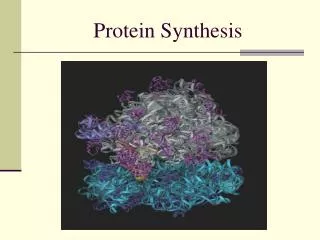
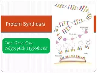
Ribosome Assembly/Structure • If individual proteins and rRNAs are mixed, functional ribosomes will assemble • Gross structures of large and small subunits are known - see Figure 30.12 • A tunnel runs through the large subunit • Growing peptide chain is thought to thread through the tunnel during protein synthesis
Eukaryotic Ribosomes • Mitochondrial and chloroplast ribosomes are quite similar to prokaryotic ribosomes, reflecting their supposed prokaryotic origin • Cytoplasmic ribosomes are larger and more complex, but many of the structural and functional properties are similar • See Table 30.6 for properties
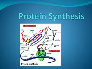
30.5 Mechanics of Protein Synthesis • All protein synthesis involves three phases: initiation, elongation, termination • Initiation involves binding of mRNA and initiator aminoacyl-tRNA to small subunit, followed by binding of large subunit • Elongation: synthesis of all peptide bonds - with tRNAs bound to acceptor (A) and peptidyl (P) sites. See Figure 30.13 • Termination occurs when "stop codon" reached
Prokaryotic Initiation • The initiator tRNA is one with a formylated methionine: f-Met-tRNAfMet • It is only used for initiation, and regular Met-tRNAmMet is used instead for Met addition • N-formyl methionine is first aa of all E.coli proteins, but this is cleaved in about half • A formyl transferase adds the formyl group (see Figure 30.15)
More Initiation • Correct registration of mRNA on ribosome requires alignment of a pyrimidine-rich sequence on 3'-end of 16S RNA with a purine-rich part of 5'-end of mRNA • The purine-rich segment - the ribosome-binding site - is known as the Shine-Dalgarno sequence (see Figure 30.17) • Initiation factor proteins, GTP, N-formyl-Met- tRNAfMet, mRNA and 30S ribosome form the 30S initiation complex
Events of Initiation • 30S subunit with IF-1 and IF-3 binds mRNA, IF-2, GTP and f-Met-tRNAfMet (Figure 30.18) • IF-2 delivers the initiator tRNA in a GTP-dependent process • Loss of the initiation factors leads to binding of 50S subunit • Note that the "acceptor site" is now poised to accept an incoming aminoacyl-tRNA
The Elongation Cycle • Elongation factor Tu will bring each aa-tRNA into the A site • Decoding center of 16S rRNA makes sure the proper aa tRNA is in the A site by direct surveillance • Peptide bond formation occurs by direct transfer of the peptidyl chain from the tRNA bearing it to the NH2 group of the new amino acid • Translocation of the one-residue-longer peptidyl tRNA to the P site to make room for the next incoming aa-tRNA at the A site.
EF-Tu-EF-Ts cycle • The elongation factors are vital to cell function, so they are present in significant quantities (EF-Tu is 5% of total protein in E. coli (Table 30.8) • EF-Tu binds aminoacyl-tRNA and GTP • Aminoacyl-tRNA binds to A site of ribosome as a complex with EF-Tu and GTP • GTP is then hydrolyzed and EF-Tu:GDP complex dissociates • EF-Ts recycles EF-Tu by exchanging GTP for GDP
Peptidyl Transferase • This is the central reaction of protein synthesis • 23S rRNA is the peptidyl transferase! • The "reaction center" of 23S rRNA is shown in Figure 30.22 - these bases are among the most highly conserved in all of biology. • Translocation of peptidyl-tRNA from the A site to the P site follows (see Figures 30.19 & 30.21 ) catalyzed by EF-G.
Peptide Chain Termination • Proteins known as "release factors" recognize the stop codon at the A site • Presence of release factors with a nonsense codon at A site transforms the peptidyl transferase into a hydrolase, which cleaves the peptidyl chain from the tRNA carrier
The Role of GTP Hydrolysis • IF-2, EF-Tu, EF-G, RF-3 are all GTP-binding proteins • Part of the G protein superfamily • Hydrolysis drives essential conformation changes • IF-2, EF-Tu, EF-G, RF-3 interact with the same site on the 50S subunit, the factor binding center
Polysomes • mRNA with several ribosomes • Polyribosomes • All protein synthesis occurs on polysomes • Procaryotes have around 10, eucaryotes fewer than 10 ribosomes