Download
1 / 60
620 likes | 1.06k Vues
Functional Human Physiology for the Exercise and Sport Sciences Chemical Messengers and the Endocrine System. Jennifer L. Doherty, MA, ATC Department of Health, Physical Education, and Recreation Florida International University. The Endocrine System. Endocrine control of cell function

E N D
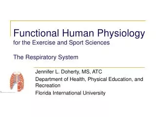
Functional Human Physiologyfor the Exercise and Sport Sciences Chemical Messengers and the Endocrine System Jennifer L. Doherty, MA, ATC Department of Health, Physical Education, and Recreation Florida International University
The Endocrine System • Endocrine control of cell function • Depends upon the secretion and action of chemical messengers or hormones • Directly linked to the autonomic nervous system • Endocrine glands • Ductless glands that release their secretory products (hormones) directly into the extra-cellular fluid. • Hormones then diffuse into capillaries and are carried throughout the body in the blood.
Specific Endocrine Glands • Primary Endocrine Glands • Hypothalamus, Pituitary, Thyroid, Parathyroid, Adrenal, Pineal glands, Thymus, Pancreas, and Gonads ( Testes and Ovaries) • Secondary Endocrine Glands • Several organs contain endocrine tissue and produce hormones • Heart, kidneys, and others • The Endocrine System is integral in Intercellular Communication
Intercellular Communication • Direct Communication through Gap Junctions • Connexins (plasma membrane proteins) link adjacent cells forming connexons • Connexons form channels that allow ions or small molecules to pass directly from one cell to another
Intercellular Communication • Indirect Communication through Chemical Messengers • Ligands (chemical messengers) bind to proteins (receptors) on the target cells • Chemical substances produced at one site cause an effect at a different site in the body. • Regulate metabolism, maintain homeostasis, and are essential for reproduction. • Binding between messenger and receptor results in a response in the target cell • Response is called Signal Transduction
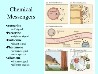
Chemical Messengers • Functional Classification (6) • Paracrines • Chemicals that communicate with neighboring cells • Autocrines • Chemicals that act on the same cell that secreted them • Neurotransmitters • Chemicals released from neurons into the interstitial fluid
Chemical Messengers • Functional Classification (6) cont. • Hormones • Chemicals released from endocrine glands • Neurohormones • Chemicals released from a special class of neurons called neurosecretory cells • Cytokines • A wide range of chemical messengers released from a variety of cells, especially WBCs
Chemical Messengers • Chemical Classification (5) • Amino Acids (Lypophobic) • Amines (Lypophobic) • Peptides (Lypophobic) • Steroids (Lypophilic) • Eicosanoids (Lypophilic)
Chemical Messengers • Lypophobic messengers • Water-soluble (hydrophilic) • Pass through the cell membrane • Function to • Open or close Channel-Linked, Enzyme-Linked, or G-Protein-Linked Receptors • Altering the permeability of the cell membrane leading to depolarization or hyperpolarization • Activate membrane bound enzymes • Activate the second messenger system (more later)
Types of lipophobic messengers • Amino Acid Messengers • Function as neurotransmitters in the central nervous system • Examples include • Glutamate, Glycine, Gamma amino butyric acid (GABA) • Most Amine Messengers • Substances derived from the amino acids • Examples include: • Catecholamines (Both norepinephrine and epinephrine), secreted by neurons as neurotransmitters or by the adrenal medulla as hormones • Peptide Messengers • Short chains of amino acids • Examples include: • Insulin, oxytocin, antidiuretic hormone (ADH)
Chemical Messengers • Lipophilic messengers • Fat loving (hydrophobic) • Do not pass through the cell membrane • Bind with receptors in the cytosol or nucleus of the target cell • Function • Control protein synthesis
Types of lipophilic messengers • Steroid Messengers • Derived from cholesterol. • Cholesterol is made up of hydrogen, oxygen and carbon molecules and is most recognizable because of its 4-ring structure. • Examples include: • Sex hormones • estrogen and testosterone • Some Amine Messengers • Derive from amino acids • Thyroid hormones • Thyroxine and triiodothyronine
Signal Transduction Mechanisms • Binding between a messenger and a receptor resulting in a response in the target cell • Produces (one or more) of four typical responses: • Changes the cell membrane permeability or membrane potential • Increases the production of proteins or regulatory molecules (enzymes) within the cell • Activates or deactivates enzymes • Increases secretory activity
Signal Transduction Mechanisms • Relationship Between Receptor Binding and the Magnitude of the Target Cell Response • Blood levels of the chemical messenger • The relative number of receptors for the chemical messenger • Affinity (strength) of the union between the messenger and receptor
Signal Transduction Mechanisms • Receptor Agonists and Antagonists • Agonists – • Ligand binds to receptor and produces biological response • Antagonists – • Ligand binds to receptor, blocking the agonist • No biological response
Signal Transduction Mechanisms • Intracellular Receptor-Mediated Responses • Lipophilic messengers affect protein synthesis of the target cell by direct gene activation • Usually involves steroid and some amine (thyroid hormones) messengers • These chemical messengers are lipid soluble. Lipids make up most of the cell membrane so they readily diffuse through the cell membrane. • Once inside the cell, the steroid messenger combines with a protein receptor usually located in the nucleus. • A messenger-receptor complex interacts with chromatin in the nucleus of cell and triggers transcription of specific genes causing production of specific mRNA for synthesis of new proteins
Signal Transduction Mechanisms • Membrane-Bound Receptor Mediated Responses • Binding of a chemical messenger or ligand to a membrane-bound receptor initiates a chain of events inside the cell that changes the cell's activity or metabolism. • Involves the amines (catecholamines), peptides, and amino acid (lipophobic) messengers • These messengers are not soluble in lipids thus, they cannot diffuse through the cell membrane and bind to intracellular receptors. • The messengers act through receptor proteins at the external surface of the cell membrane and depend on second messengers inside the cell to mediate the cellular response to the chemical messenger.
Signal Transduction Mechanisms • G-Protein Linked Receptors • Involves the intracellular enzymes Cyclic AMP, Adenylate Cyclase, and Phosphodiesterase • Cyclic adenosine monophosphate (cAMP) • The best known second messenger • Adenylate cylase • Cyclic AMP is synthesized in the cell from ATP via the action of an enzyme attached to the inner surface of the plasma membrane, adenylate cyclase. • Phosphodiesterase • cAMP is inactivated by another enzyme present in the cell, phospho-diesterase.
G-Protein second messenger systems: • The hormone is the first messenger and it binds to receptor on the cell membrane, usually a G protein. • The G-protein activates adenylate cyclase that generates cAMP from intracellular ATP (G protein is a transducer) • cAMP is the second messenger • It initiates a cascade of reactions by activating protein kinases which phosphorylate millions of proteins/enzymes, producing an amplification effect. • Phosphorylation activates some proteins, but deactivates others. It is like an on/off switch thus, cAMP can lead to many different physiological responses. • Different cells contain different proteins so that cAMP is able to produce different effects in different cells often with several different actions in one cell at the same time. • cAMP is rapidly degraded by phosphodiesterase. This turns off the cellular response, unless new hormone molecules continue to bind to the membrane-bound receptor. There are other known second messengers, cAMP is the best understood.
Signal Amplification in Chemical Messenger Systems • The ability of small changes in the concentration of a chemical messenger to elicit marked responses in target cells • A single kinase enzyme can catalyze thousands of reactions • As the reaction cascades through enzymes, one intermediately after another, the number of product molecules increases dramatically. • For example, one kinase enzyme can activate many G-proteins producing thousands of cAMP molecules
Specific Endocrine Glands and their Hormones • Control of Hormone Levels • Negative feedback • The concentration of each hormone in the body fluid is regulated precisely by negative feedback systems. • In a negative feedback system, a gland is sensitive to the concentration of a substances it regulates. • When the concentration of the regulated substance reaches a certain concentration, it inhibits the gland. As the gland secretes less hormone, the controlled substance also decreases. • Feedback systems occur when a hormone level or its effect is fed back to the gland. • The endocrine gland then responds in a manner that will return the system to homeostasis. • For example: • Increased blood glucose concentrations stimulate insulin secretion by the pancreas. Insulin stimulates glucose uptake by cells decreasing the blood glucose concentration and inhibiting insulin secretion.
Three types of stimuli affect endocrine glands • Hormonal stimuli • Produce responses in the same or other endocrine glands. • For example, the hypothalamus secretes releasing hormones or inhibiting hormones to the anterior pituitary gland. • Increased release of particular anterior pituitary hormone into blood stream tells the hypothalamus to decrease secretion of releasing hormones. • Decreased secretion of the releasing hormones decreases the activity of the anterior pituitary. • Humoral stimuli • Refers to blood and other body fluids. This term refers to chemical changes in the blood that can influence endocrine gland activity. • For example, changes in blood glucose concentration produces changes in insulin secretion by the pancreas. • Neural stimuli • Long-distance communication via the nervous and endocrine systems • Some endocrine glands secrete in response to neural stimuli or nerve control. • Neural stimuli results from nerve fibers signaling hormonal release from a gland. • For example, the sympathetic nervous system stimulates the adrenal medulla to release catecholamines during periods of stress such as, exercise.
Primary Endocrine Glands • Main function is to secrete hormones • Hypothalamus and Pituitary Gland • Hypothalamus • Master control of one of the most important endocrine glands, the pituitary. • It contains centers for control of body temperature, appetite, thirst, blood nutrient concentrations, sexual behavior, and emotional state. • Functions as an important link between the nervous and endocrine systems.
Anterior Pituitary Gland • Controlled by the hypothalamus. • The hypothalamus controls the secretory activity of the anterior pituitary by producing releasing hormones (RH) and inhibiting hormones (IH). • Releasing hormones produced by the hypothalamus cause hormone release from the anterior pituitary. • Inhibiting hormones produced by the hypothalamus slow or suppress the release of certain anterior pituitary hormones.
Hypothalamic control is regulated via negative feedback • When blood concentration of a particular hormone rises to a certain level, the hypothalamus either decreases production of releasing hormone or produces inhibiting hormone. • Hypophyseal portal veins • Connect the hypothalamus to the anterior pituitary • Transports hypothalamic releasing and inhibiting hormones to the anterior pituitary gland.
Anterior Pituitary Hormones • Growth Hormone (GH) • Protein hormone with target tissues throughout the body (bones and muscles being the primary target cells). • General effect of growth hormone • Promote cell growth and division (anabolic effect) by stimulating the uptake of amino acids and protein synthesis, while slowing protein catabolism. • Increases the growth rate of skeleton and skeletal muscles during childhood and adolescence. • In adults, growth hormone helps maintain muscle and bone size and promote tissue repair. It affects growth in target cells indirectly, through proteins called somatomedins.
Prolactin • A protein hormone that initiates and maintains milk secretion by the mammary glands in women. • Regulated by • Prolactin inhibiting hormone (PIH) from the hypothalamus • Prolactin-releasing hormone (PRH) also from the hypothalamus • Normally, prolactin inhibiting hormone predominates over prolactin-releasing hormone (PRH) which suppresses milk production. Prolactin release-inhibiting factor from the hypothalamus restrains secretion of prolactin, while prolactin-releasing factor promotes its secretion.
Melanocyte stimulating hormone (MSH) • The exact role in humans is unknown. • Tropic hormones • Regulate the activity of other endocrine glands. • There are no hypothalamic inhibiting factors associated with the tropic hormones, only releasing hormones. • Thyroid stimulating hormone (TSH) or thyrotropin • Stimulates normal development and secretory activity of the thyroid gland. • Stmulates synthesis and secretion of thyroid hormones. • Adrenocorticotropic hormone (ACTH) • Target organ as the adrenal cortex. • Stimulates release of corticosteroid hormones, especially cortisol from adrenal cortex. • Release is stimulated by corticotropin releasing hormone (CRH) from the hypothalamus.
Gonadotropins • Hormones that stimulate the hormonal functions of the gonads (ovaries and testes). • Follicle stimulating hormone (FSH) • Females • Stimulates the development of the follicle and egg in the ovaries and stimulates the follicles to secrete estrogen (female sex hormone). I • Males • This hormone known as interstitial cell stimulating hormone (ICSH) • Stimulates the interstitial cells of the testes to release testosterone and stimulates sperm cell production. • Luteinizing hormone (LH) • Females • Stimulates the maturation of the egg and its release from the ovary. This includes ovulation or expulsion of the egg from the follicle, development of the corpus luteum, release of the ovarian hormones estrogen and progesterone. • Males • Effects are not clinically important
Posterior pituitary gland hormones • Oxytocin • A protein hormone with two target tissues, the uterus and breast. • During childbirth, it stimulates the smooth muscle contractions in the walls of uterus. Also, stimulates ejection of milk from breast glands during lactation in response to the mechanical stimulation from suckling infant. • Example of a positive feedback mechanism • Antidiuretic hormone (ADH) • Also called vasopressin • Effects of antidiuretic hormone • Decrease urine volume produced by the kidney (an antidiuretic) resulting in more fluid returned to the blood. • This increased blood volume produces increased blood pressure. • Also stimulates smooth muscle contraction in arterioles (small blood vessels) increasing blood pressure.
Thyroid Hormones • Thyroxine (T4 ) • Accounts for almost 95% of circulating thyroid hormone, although T3 is the more active form • Triiodothyronine (T3) • T3 and T4 function to: • Stimulate cellular metabolism • Increased production of oxidative enzymes and of Na+/K+ pumps • Increased basal metabolic rate and metabolic heat production • Increased heart rate and force of contraction • Increased blood pressure from up-regulation of catecholamine receptors • The thyroid hormones are important in normal tissue growth and development, maturation of the nervous system
Calcitonin • A peptide hormone produced the thyroid gland. • Functions • Lower the blood calcium levels by inhibiting osteoclasts and stimulating osteoblasts. • Bone-sparing effect • Secretion of calcitonin is stimulated when blood calcium concentration is high such as immediately after a meal. • Calcitonin works opposite (antagonist) of the parathyroid hormone in regulation of blood calcium levels
Parathyroid hormone (PTH) • A protein hormone secreted by the parathyroid glands • Functions as a second messenger to • Increase blood [calcium] and decrease blood [phosphate] by stimulating osteoclast activity and increasing bone resorption thus increasing blood calcium levels. • Release is stimulated by decreased blood calcium levels • Parathyroid hormone is the single most important regulator of calcium levels in adult humans. • Important for normal transmission of nerve impulses, muscle contraction, and blood clotting. • Abnormalities of blood calcium levels result in depression of the nervous system, abnormal reflexes, weak muscles, twitches, and formation of kidney stones
Hormones of the Adrenal Cortex • The adrenal cortex can be divided into three zones that produce different types of corticosteroid hormones. • From superficial to deep: • Zona glomerulosa • produces aldosterone, a mineralocorticoid • Zona fasciculata • produces cortisol, the most abundant glucocorticoid • Zona reticularis • produces the adrenal sex hormones, primarily the androgens and estrogens or the gonadocorticoids.
Corticosteroids • The collective term for the steroid hormones secreted from the adrenal cortex that are essential for life. • The corticosteroids include the mineralcorticoids, glucocorticoids, and the gonadocorticoids. • Mineralcorticoid • Aldosterone • Functions to maintain water and electrolyte homeostasis, especially blood sodium and potassium levels by stimulating sodium reabsorption (conservation) and potassium excretion by the kidneys. • Sodium is returned to the blood (with water) producing decreased urine volume, increased blood volume, and increased blood pressure.