10.
3. 4. central sulcus cerebellum cortex fissure frontal lobe gyrus lateral sulcus medulla oblongata occipital lobe parietal lobe parieto -occipital sulcus pons postcentral gyrus precentral gyrus sulcus temporal lobe transverse cerebral fissure white matter. 2. 5. 1. 6. 7.

10.
E N D
Presentation Transcript
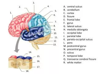
3. 4. central sulcus cerebellum cortex fissure frontal lobe gyrus lateral sulcus medulla oblongata occipital lobe parietal lobe parieto-occipital sulcus pons postcentralgyrus precentralgyrus sulcus temporal lobe transverse cerebral fissure white matter 2. 5. 1. 6. 7. 8. 9. 10. 11. 12. 13. 14. 15. 16. 17. 18.
auditory association area complex problem solving frontal eye field gustatory cortex prefrontal cortex premotor cortex primary auditory cortex primary motor area primary somatosensory cortex primary visual cortex somatosensory association area visual association area Wernicke’s area working memory, recall 7. 1. 8. 2. 3. 9. 10. 4. 11. 12. 5. 13. 6. 14.
anterior commissure frontal lobe hypothalamus interthalmic adhesion interventricular foramen mammillary body medulla oblongata optic chiasma pituitary gland pons septum pellucidum spinal cord temporal lobe 1. 2. 3. 4. 5. 6. 7. 8. 9. 10. 11. 12. 13.
arbor vitae cerebellum cerebral aqueduct choroid plexus choroid plexus corpora quadrigemina corpus callosum fornix fourth ventricle midbrain occipital lobe parietal lobe pineal gland posterior commissure thalamus 1. 2. 3. 4. 5. 6. 7. 8. 9. 15. 10. 11. 12. 13. 14.
1. 7. 2. arachnoid mater arachnoid villus dura mater falxcerebri meningeal periosteal pia mater subarachnoid space subdural space superior sagittal sinus 3. 8. 4. 9. 5. 10. 6.
arachnoid villus central canal cerebral aqueduct choroid plexus confluence of sinuses fourth ventricle intraventricular foramen lateral aperture median aperture straight sinus subarachnoid space superior sagittal sinus tentorium cerebelli third ventricle 1. 7. 2. 8. 9. 3. 4. 10. 5. 11. 12. 13. 6. 14.
1. 5. 2. 6. arachnoid dorsal root ganglion dura mater epidural space pia mater subarachnoid space subdural space 3. 7. 4.
1. anterior funiculus anterior horn anterior median fissure arachnoid central canal dorsal horn dorsal root dorsal root ganglion gray commisure lateral funiculus lateral horn pia mater posterior funiculus posterior median sulcus spinal mater spinal nerve ventral root 2. 11. 3. 12. 4. 13. 5. 14. 15. 6. 16. 17. 7. 8. 9. 10.