Jaundice – For the practitioners
710 likes | 2.45k Vues
Dr.R.V.S.N.Sarma., M.D., M.Sc., Consultant Physician & Chest Specialist. A Lucid Understanding of. Jaundice – For the practitioners. Jaundice – Classification. Normal Serum Bilirubin (SB) is 0.3 to 1.0 mg% Jaundice is increased levels of SB > 1.0 mg%

Jaundice – For the practitioners
E N D
Presentation Transcript
Dr.R.V.S.N.Sarma., M.D., M.Sc., Consultant Physician & Chest Specialist A Lucid Understanding of Jaundice – For the practitioners
Jaundice – Classification www.drsarma.in • Normal Serum Bilirubin (SB) is 0.3 to 1.0 mg% • Jaundice is increased levels of SB > 1.0 mg% • Over production of Bilirubin (Hemolytic) • From hemolysis of RBC • Lysis of RBC precursors – Ineffective erythropoesis • Impaired hepatic function (Hepatitic) • Hepatocellular dysfunction in handling bilirubin • Uptake, Metabolism and Excretion of bilirubin • Obstruction to bile flow (Obstructive) • Intrahepatic cholestasis • Extrahepatic Obstruction (Surgical Jaundice)
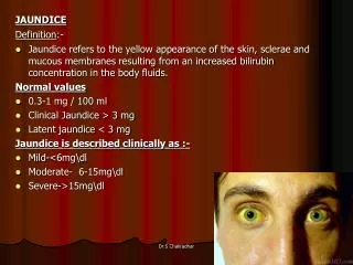
Clinical Aspects of Jaundice www.drsarma.in • Clinically detectable if SB is >2.0 mg% • With edema and dark skin – Jaundice is masked • What is special about the sclera ? – Rich Elastin • Darkening of the urine – Differential Diagnosis • Skin discoloration – Yellowish, - Carotinemia – Eyes N • Mucosa – hard palate (in dark skinned) • Greenish hue of skin and sclera - due Biliverdin – indicates long standing jaundice • Generalized Pruritus – Obstructive Jaundice – Why ?
Clinical History – Imp clues www.drsarma.in • Duration of jaundice – Acute / Chronic • Abdominal pain v/s painless jaundice • Fever – Viral / bacteria /sepsis • Arthralgia, rash, glands; Pruritus - obstructive • Appetite – Hepatocellular / Malignancy • Weight loss – Malignancy – CAH • Colour of stools –chalky white –obstructive • Family history – Hemolytic – Inherited dis. • H/o transfusion, promiscuity, IDU • Alcohol abuse, Medications – INH, EM, Largactil
Coloured Urine – Differ. Diagnosis www.drsarma.in • Bilirubin in urine due to Jaundice (CB) • Concentrated urine in dehydration • Fluid deprivation syndromes • Sulfasalazine use – for Ulcerative colitis • Rifampicin, Pyridium and Thiamine use • Red urine – Porphyria, • Hemoglobin & Myoglobinuria, Hematuria • Dark black urine in Ochranosis - HGA • Melanin excretion from Melanoma • Red sweat in Clofazamine, Rifampicin
Fate of Senescent RBC • RBC life span in blood stream is 90-120 days • Old RBCs are phagocytosed and/or lysed • Lysis occurs extravascularly in the RE system subsequent to RBC phagocytosis • Intravascular Hemolysis of young RBC • This is due to hemolytic diseases of RBC www.drsarma.in
The Hepatobiliary & Portal System Hepatobiliary Tree Portal Circulation www.drsarma.in
E V Pathway for RBC Scavanging Liver, Spleen & Bone marrow Phagocytosis & Lysis Hemoglobin Heme Bilirubin Globin Fe2+ Amino acids Through Liver Excreted Amino acid pool www.drsarma.in
Bilirubin Handling www.drsarma.in
Bilirubin Metabolism - Summary www.drsarma.in
Bilirubin – And its nature www.drsarma.in
Bilirubin in the Liver Cell www.drsarma.in
Bilirubin in Liver Cell - Schematic www.drsarma.in
Blood ALT ALT ALT ALT ALT ALT ALT ALT ALT ALT ALT ALT ALT ALT ALT ALT ALT ALT ALT ALT ALT ALT ALT ALT ALT ALT ALT AlkP AlkP AlkP AlkP AlkP AlkP AlkP AlkP AlkP AlkP AlkP AlkP AlkP AlkP AlkP Bile
Blood ALT ALT ALT ALT ALT ALT ALT ALT ALT ALT ALT ALT ALT ALT ALT ALT ALT ALT ALT ALT ALT ALT ALT ALT ALT ALT ALT AlkP AlkP AlkP AlkP AlkP AlkP AlkP AlkP AlkP AlkP AlkP AlkP X Bile
Bilirubin in the Intestine www.drsarma.in
Bilirubin handling in Kidney www.drsarma.in
An Approach to Jaundice • Is it isolated elevation of serum bilirubin ? • If so, is the↑unconjugated or conjugated fraction? • Is it accompanied by other liver test abnormalities ? • Is the disorder hepatocellular or cholestatic? • If cholestatic, is it intra- or extrahepatic? • These can be answered with a thoughtful • History and physical examination • Interpretation of laboratory tests and • Radiological tests and procedures. www.drsarma.in
Bilirubin testing www.drsarma.in Van den Berg Reaction SB + SAA Diazo compound formed Diazo is chromogenic – Colourimerty Reaction in H2O medium – Direct – CB Reaction in ethnol medium – Indirect Indirect includes CB and UCB = Total B Time is the essence of the direct test Foam test, Ictotest for urine – CB only
Normal values for LFT www.drsarma.in
Lab Diagnosis of Jaundice – D.D www.drsarma.in
Liver Function Tests (LFT) www.drsarma.in
Utility of Liver Function Tests www.drsarma.in
Non Hepatic causes of abnormal LFT www.drsarma.in
Algorithmic approach for Jaundice How to clinically evaluate the patient ? What tests will help us in D.D ? What imaging modalities will be useful ? How to monitor the progress ? www.drsarma.in
First Step www.drsarma.in
Second Step : If SB > 1.0 mg www.drsarma.in
↑ in Unconjugated Bilirubin www.drsarma.in
Third Step : If CSB is increased www.drsarma.in
Fourth Step : Hepatocellular www.drsarma.in
What imaging we need www.drsarma.in Ultrasonography – 98% Sp, 90% Sen. For GB stones USG better than CT For duct stones –only 40% seen in USG PTC – Extrahepatic obstr. – drainage ERCP – Distal biliary obstruction Dx.Rx. MRCP – Most useful for duct stones
Neonatal Jaundice www.drsarma.in Neonatal jaundice is common 50% healthy term infants Re-emergence of kernicterus In utero bilirubin is handled by placenta and mother’s liver After birth, neonate to has cope with increase in bilirubin production and the immature liver cannot handle for a few days
Treatment options for neonatal jaundice www.drsarma.in
Basis of photo therapy ? www.drsarma.in UCB is not water soluble in its form Blue light confrontational change in UBG Its Photo Isomers are water soluble Blue light converts the UCG into its photo isomers The soluble photo isomers pass through the Glomerular filter and get excreted Thus conjugation in liver is by passed.
Post hepatic Obstructive Jaundice • Painful v/s painless • Obstruction can be • Luminal (stone) • Stricture (benign v/s cholangiocarcinoma) • Extra luminal pancreatic cancer, Sec. lymph nodes • Investigate & treat with • Radiology (US, CT, MRCP) • ERCP / PTC www.drsarma.in
Chronic Liver Disease (CLD) • Alcoholic Liver (ALD) • Chronic viral hepatitis • Hepatitis B • Hepatitis C • Autoimmune liver disease: • Autoimmune hepatitis • Primary Biliary Cirrhosis (PBC) • Inherited conditions • Haemochromatosis • Wilson’s Disease • Alpha1-Antitrypsin Deficiency (AATD) • Non-alcoholic steato-hepatitis (NASH) • Budd-Chiari syndrome • Cryptogenic www.drsarma.in
Hepato toxic drugs www.drsarma.in
Acute Cholecystitis GB wall is thickened and striated. Courtesy of Udo Schmiedl, M.D. www.drsarma.in
Causes of Cholestatic Jaundice www.drsarma.in
Drugs causing Cholestasis www.drsarma.in
Complications of CLD • Portal hypertension • Varices • Ascites • Hypersplenism • Synthetic dysfunction • Coagulopathy • Encephalopathy • Immunodeficiency • Malnutrition • Hepato-cellular carcinoma www.drsarma.in
KF Ring of Periphery of Iris Courtesy of Robert L. Carithers, Jr., M.D.
Magnetic Resonance Cholangio-Pancreatography (MRCP) Two stones in the common bile duct Courtesy of UdoSchmiedl, M.D.
Retrograde Cholangiogram - ERCP Bile leak from the cystic duct after cholecystectomy Courtesy of Michael Kimmey, M.D.
Retrograde Cholangiogram - ERCP Primary sclerosing cholangitis (PSC) with stricture due to cholangiocarcinoma. Courtesy of Robert L. Carithers, Jr., M.D.
Retrograde Cholangiogram - ERCP Irregular dilation of intrahepatic and extrahepatic ducts. Courtesy of Charles Rohrmann, M.D.
Primary Sclerosing Cholangitis Narrowed abnormal intra-heptic bile ducts. Normal Extra hepatic BD
Alcoholic Cirrhosis of Liver The cut surface of a autopsy liver of a patient with alcoholic cirrhosis - multiple small nodules and diffuse scarring. Courtesy of Robert L. Carithers, Jr., M.D.
CT Abdomen A large mass with a hepatoma. Courtesy of Udo Schmiedl, M.D.