Download
1 / 27
270 likes | 520 Vues
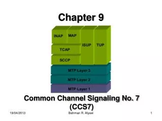
TGF- Signaling in Stem Cells & Cancer. Helen Hwang 4.22.2009. L Mishra, R Derynck, B Mishra Science: October 2005. Germ layers eventually give rise to all of an animal’s tissues and organs. fertilization zygote blastula – ball gastrula – 3 germ layers organogenesis.

E N D
TGF- Signaling in Stem Cells & Cancer Helen Hwang4.22.2009 L Mishra, R Derynck, B Mishra Science: October 2005
Germ layers eventually give rise to all of an animal’s tissues and organs. • fertilization • zygote • blastula – ball • gastrula – 3 germ layers • organogenesis
Embryonic stem cells develop into multiple functional cell lineages. ES differentiation - process where less specialized more specialized cell from pluripotent progenitor functional cells hematopoietic (bone marrow) RBC, WBC, platelets mesenchymal (bone marrow) stromal, fat, bone epithelial skin neuronal Cell signaling controls differentiation via growth factors ES growing on fibroblasts
TGF- signaling underlies progression of differentiation. • maintains undifferentiated state • initiates differentiation • specifies germ layer differentiation • depends on: • stage of target cell • local environment • identity/dosage of ligand http://stemcells.nih.gov, 2001
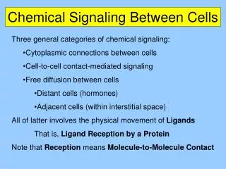
TGF- family “transforming/tumor growth factor” includes 30 structurally related growth factors TGF-s activins BMPs (bone morphogenetic protein) myostatin 2 types of serine-tyrosine receptors (type I & II) functionally: promote or inhibit cell proliferation promotes apoptosis differentiation
early: • BMPs • inhibits differentiation • after progenitors established: • promotes differentiation (ganglions, olfactory neurons) • accelerates differentiation & lineage commitment of precursor cells • fully differentiated: • inhibits growth of normal glial cells (tumors) Differentiation of neural stem cells involves TGF-
early specification: • TGF-inhibits early multipotent hematopoietic stem cells (in vitro) • BMPs • promote specification • differentiation • proliferation • progression along lineage • myeloid: promoted by Smad 7 • lymphoid: inhibited by Smad7 • dependent on exogenous factors (other growth factors, cross-talk) TGF- signaling in hematopoietic stem cells is complex
TGF- Signaling in mesenchymal stem cells • promotes specification • allow mesenchymal cells from one lineage to switch to another lineage (pre-adipocytes osteoblasts) • inhibits progression and maturation of myoblasts (myostatin), osteoblasts (BMPs), and adipocytes (myostatin) • TGF- expression are activated in response to injury (wound repair)
TGF- Signaling in gastrointestinal tissues & cancer • tumor supressors • inhibits cell growth / cancer in gut epithelial • inactivation of any signaling GI tumor • BMP signalling • suppresses Wnt signaling effects limits cell renewal • mutations in R & Smad4 intestinal polyposis or Cowden disease • TGF- signaling (Smad2, Smad3, ELF) all necessary for proper liver and biliary system development • knockouts – hepatocellular carcinoma polyposis hepatocellular carcinoma
Conclusion • TGF- is a key regulator in ES differentiation and progression of cell lineage of progenitor cells • Environmental factors and cross-talk b/t pathways could affect differentiation • When TGF- pathway is deregulated, depending on the stage impaired differentiation and may become cancerous!
Smad3-dependent translocation of -catenin is required for TGF-b1-induced proliferation of bone marrow-derived adult human mesenchymal stem cells Hongyan Jian, Xing Shen, Irwin Liu Mikhail Semenov, Xi He, Xiao-Fan WangGenes & Development, 2006.
Mesenchymal stem cells (MSC) differentiate into bone, muscle, tendon, & adipose. derived from bone marrow TGF- involved in wound repair
Question: What kind of regulatory mechanisms underlie the renewal and differentiation of MSC?
TGF-1 induces nuclear translocation of -catenin independently of the Wnt signaling pathway • incubated MSC with Wnt3A (6h) or TGF- medium (2h) • measured presence of -catenin via Western blotting • detected nucleus translocation for both
TGF-1 induces nuclear translocation of -catenin independently of the Wnt signaling pathway with immunofluorescence imaging, nuclear staining of endogenous -catenin increased in MSC 1h after treatment with TGF-1 Hoechst dye – stains DNA (visualize nuclei or mitochondria)
-catenin nuclear translocation is associated with certain cell types (MSCs) • Are TGF- 1 effects only associated with certain cellular contexts? • take Madin-Darby canine kidney epithelial cells (shown), and human fibroblasts, and human melanocytes • TGF- and Wnt3A treatment • TGF-1 did not induce -catenin accumulation although Wnt3A treatment did.
TGF-1’s translocation activity is not mediated by Wnt proteins. • Is -catenin translocation a consequence of TGF-1 induced Wnt production & action? • MSCs were pretreated with protein translation inhibitor (cyclohexmide) for 1 hr • treat with TGF- 1 for 2 hrs • detect presence of -catenin, -tubulin, lamin • CHX had no effect on TGF- 1’s effect
b-catenin is dependent on TGF-b type 1 receptor • Treat MSC with SD208 (kinase inhibitor of TGF- type 1 receptor) • apply TGF-1 • Smad2 phosphorylation is blocked and -catenin translocation blocked
SMads are directly involved in b-catenin translocation (via Smad-KDs) • introduce Smad specific siRNA (Smad3 protein reduced by >90%) • apply TGF-1 in MSC • examine -catenin nuclear translocation • -catenin protein is barely detectable in Smad-KDs
TGF-1 and nuclear -catenin both increase proliferation • b-catenin mutants formed via retroviral infection • full transcriptional activity • alanine instead of serine p sites so unable to degrade (via ubiquitin) • treat with TGF- 1 or untreated in control or mutant b-catenin/vector • treat with H3-thymidine • measure relative proliferation of human MSCs • TGF- 1 & -catenin mutants both have increased relative proliferative activity
TGF-B1 and nuclear b-catenin are both anti-osteogenic • osteogenic assay – measure alkaline phosphate activity • culture MSC’s in osteogenic (OS) medium • treat in presence/absense of TGF- or look at -catenin mutants • TGF-b and b-catenin inhibits the osteogenic effect of the OS medium on MSCs. • perhaps direct correlation between -catenin and TGF- 1
TGF-b1 mediates proliferative effect on MSCs via b-catenin translocation • LEF1 – transcription factor that complexes with -catenin that translocates into nucleus via HMG box (where LEF1 & Smad3 interacts) • LEF1ΔC - a mutant of LEF1 that can still complex with -catenin in cytoplasm, but cannot translocate into nucleus (unable to associate with Smad3) • In LEF1ΔC, TGF-1 did not induce proliferation. • In LEF1ΔC, TGF-1 did not inhibit osteogenic differentiation. • -catenin is required for TGF- to exert some of its biological effects on MSCs.
Some unanswered questions • Interactions exist in other cells, but why does SMAD3 only work in MSCs? • Opposite physiological effects seen in human MSCs as opposed to other stem-cell types. Why? • How do TCF/LEF transcription factors participate in the proliferative response seen? • What kind of downstream mechanisms exist after SMAD3 but before -catenin in the Wnt and TGF- pathway?
Conclusions • Smad3 plays a role in the translocation of b-catenin into nucleus through a process initiated by TGF-b1 • This is a novel signaling pathway found only in MSCs