Download
1 / 19
200 likes | 534 Vues
LAB: Sensory Illusions E xploration. What? How? Why?. Visual illusions. Visual illusions are often the hardest to overcome, even when we know that our senses are being deceived. This may be due to the overwhelming amount of neural tissue and activity associated with the sense of vision.

E N D
LAB: Sensory Illusions Exploration What? How? Why?
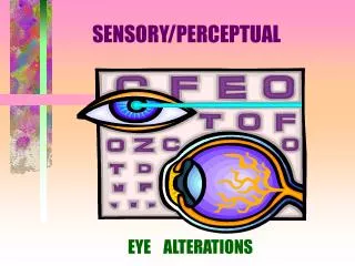
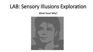
Visual illusions • Visual illusions are often the hardest to overcome, even when we know that our senses are being deceived. • This may be due to the overwhelming amount of neural tissue and activity associated with the sense of vision. • Perhaps as much as 75% of the sensory input we receive is visual and 25% of the processing power of our brains is associated with this sense. • Not surprisingly, more is known about the functioning of the vertebrate eye than any other sensory structure.
Hermann grid • The retina is partially composed of many small nerves, called rods, which receive and transduce radiant energy into electrochemical action potential. • The rods are arranged in rows on the inside of your retina and are responsible for light and darkvision. • If light strikes a rod, the energy is transduced into an electrochemical signal and the visual cortex of the brain produces a “white” image. If the photoreceptor is not illuminated, no signal is sent and the brain perceives “black” at that location in the retina.
Hermann grid (cont’d) • Illumination a single rod produces a large white response in the visual centers of the brain. However, when you add illumination to the adjacent rods, the response in the brain from the original rod is reduced. • In other words, illumination of photoreceptors "inhibits" of firing of neighboring receptors. This effect is called lateralinhibition because it is transmitted laterally, across the retina, in a structure called the lateralplexus.
Color Afterimages • When radiant energy strikes the retina, it may be transduced by color photoreceptors called cones. • The output of these receptors is converted in the retina in an opponencyprocess. In this process the output of the cones is sent via the network of over one million nerve fibers comprising the opticnerve. • The optic nerve encodes color in three separate channels, one for intensity and two for color. One set of neurons encodes black-whitedifferences, corresponding to intensity or luminance differences in the image (similar to looking at a scene through a black-and-white video camera).
Color Afterimages (cont’d) • Another set of neurons responds to red-greencolordifferences and a third set responds to yellow-bluedifferences. • For instance, a red-green cell would increase its activity as a result of stimulation with red (R) light and would decrease its activity in response to green (G) light. It can be said to signal +R-G. Other cells signal the opposite, that is the presence of green and the absence of red (+G-R). As such, one could say that green is the opponent of red. • In the third pathway, blue light would signal +B-Y while yellow would signal +Y-B. What we perceive as “color” is really composite of the relative activity in these three pathways of the optic nerve.
Color Afterimages (cont’d) • All three photoreceptive cones utilize a chemical called photopsin. • When photopsin is struck by light, it undergoes a change in its 3D form which in turn triggers a series of chemical messengers that ultimately produce electrochemical stimulation of the visual centers in the brain. • Rods have a similar chemical called rhodopsin for black/white colors.
Color Afterimages (cont’d) • Once photopsin has changed its 3D form, it may take as long as 45 minutes for it to change back to its original form. During that period of time, the photopsin is said to be photobleached and it sends a constant stimulus to the brain. • Eventually, the brain becomes desensitized to the constant stimulation from that color photoreceptor and the brain does not see that color. However, due to the opponency process, the complimentarycolor appears as an “afterimage”.
Auditory Illusions • Auditory illusions (i.e. hearing) may not utilize as much of the brain’s processing as vision. That said, audition (along with tactile sense) serves a critical role in perception of one’s environment as well as risk-avoidance. • While the brain may devote more space to the processing of visual stimuli, the perception of hearing is actually fivetimesfaster (0.05 secs v. 0.25 seconds). • The normal, adult human ear can hear anywhere between 20-20,000 Hz.
Auditory Illusions (cont’d) • The transduction of sound waves into electrochemical messages requires at least three changes of energy in the outer, middle and innerears. • Ultimately, it is the movement of the specialized hairs in the basilarmembrane of the cochlea that sends action potentials to the brain via the auditory nerve. • Audition (hearing), along with vision, has the one of the highest degrees of precision and accuracy of our sense. That said, the ear can be “fooled” just like any other sense through the right presentation of tones.
Tactile Illusions • Despite the fact that our skin is the largest and heaviest organ in our body, containing millions of tactile receptors, touch remains one of the more mysterious and complex of our senses. • This is due to the polymodal nature of tactile sensation; many of these receptors can receive multiple forms of stimuli and combine their input to create unique net effects such as temperature sensation, pressure, wetness and itchiness. • The skin contains at least three major types of receptors: • Merkel Receptors- light touch • Pacinian Corpuscles – deep touch & pressure • Ruffini Corpuscles– stretching and temperature sensation
Tactile Illusions (cont’d) • Both Merkel and Pacinian Corpuscles are capable of adaptation, the phenomenon that we have seen before in which the nervous system compensates for constant sensory input. • Ruffini Corpuscles, however, do not readily undergo adaptation. This is because Ruffini Sensors are examples of nociceptors, specialized receptors that communicate pain directly to the central nervous system.
Olfactory & Gustatory Illusions • While the previous three senses (vision, hearing and touch) have clearly defined pathways that tend not to interact with other senses, the senses of smell and taste are intimately connected in the concept of flavor. • Flavor is a combination of taste (the electrochemical impulses from the tongue), olfaction (electrochemical impulses from the nasal passages) and other stimuli from the trigeminalnerve (greatest sensory nerve of face/head, involved in mastication). • Both senses employ similar forms of chemoreceptors: the gustatorypapillae of the tongue and the olfactoryreceptorneurons (ORNs) of the nasal passages are responsive to only one specific molecule each. While we don’t eat through our nasal passages, chewing does allow for the passage of airborne molecules into the nasal cavity.
Olfactory & Gustatory Illusions • Our tongues are the location of appx. 10,000 pear shaped modified epithelial cells that house a chemical called gusductin. • Changes in the shape of gusductin cause electrochemical impulses to be sent to the brain. Likewise, the ORNs contain a protein called GOLF that similarly triggers a second wave of chemical messengers. • These receptive neurons of taste and smell share many similarities with the rods and cones of the retina, yet they differ in how the receptive cells of the retina do not rely on interaction with other sensory input. • In other words, you don’t need to hear to have well-developed vision but anosmia (loss of smell) will greatly diminish one’s sense of taste.
Colorblindness • There are four kinds of color vision:Regular vision is Trichromatic - it uses all three color receptors (red/green/blue). In practice, the cone cells in your eyes are called L, M and S (for long, medium and short wavelength reception), but the colors they 'see' are closer to Yellow, Green and Blue. • The wavelengths they pick up are vastly overlapping, so green light hits all three in varying degrees (and all wavelengths hit your rod cells).
Colorblindness • People with AnomalousTrichromatic vision use all three color receptors but reception of one pigment is misaligned. • Protanomaly: reduced red sensitivity. • Deuteranomaly: reduced green sensitivity. • Tritanomaly: reduced blue sensitivity. • People with Dichromatic vision use only 2 of the 3 visual pigments - red, green or blue is missing. • Protanopia: unable to receive red. • Deuteranopia: unable to receive green. • Tritanopia: unable to receive blue.
Colorblindness • Monochromat (Achromatopsia) • People with Monochromatic vision can only see one color, so their vision contains no 'color'. • TypicalMonochromatic: unable to combine colors. Fully grayscale. • Also known as Rod Monochromat • AtypicalMonochromatic: very low color recognition. • Also known as Cone Monochromat
Colorblindness • Some fish, birds, and insects are tetrachromats • Ultraviolet sensors in addition to RGB