Cell Division
210 likes | 307 Vues
Explore the intricacies of cell division functions in growth, repair, and reproduction. Learn about mitosis and meiosis, chromosome structure, cell cycle phases, and the differences between normal and cancerous cells. Discover the stages of meiosis and the significance of each phase.

Cell Division
E N D
Presentation Transcript
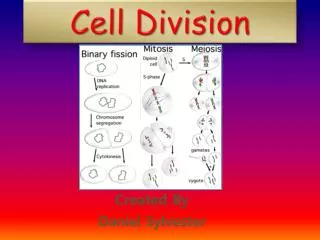
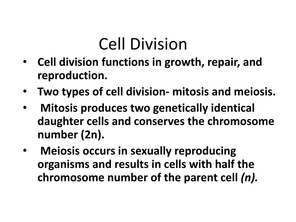
Cell Division Cell division functions in growth, repair, and reproduction. Two types of cell division- mitosis and meiosis. Mitosis produces two genetically identical daughter cells and conserves the chromosome number (2n). Meiosis occurs in sexually reproducing organisms and results in cells with half the chromosome number of the parent cell (n).
Chromosomes • A chromosome consists' of a highly coiled and condensed strand of DNA. • A replicated chromosome consists of two sister chromatids, where one is an exact copy of the other. • The centromere is a specialized region that holds the two chromatids together. • The kinetochore is a disc-shaped protein on the centromere that attaches the chromatid to the mitotic spindle during cell division.
Two factors limit cell size and promote cell division • ratio of the volume of a cell to the surface area • capacity of the nucleus to control the entire cell The nucleus must be able to provide enough information to produce adequate quantities to meet the cell's needs.
Phases of the Cell Cycle • Five major phases: G1, S, and G2(which together make up interphase), mitosis, and cytokinesis.
Interphase • Consists of G1, S, and G2. • The G1is a period of intense growth and biochemical activity. • Sis the synthesis or replication of DNA. • G2 is the phase when the cell continues to grow and to complete preparations for cell division. • More than 90 percent of the life of a cell is spent in interphase. • During interphase chromatin is threadlike, not condensed. • Within the nucleus are one or more nucleoli. • A centrosome, consisting of two centrioles, can be seen in the cytoplasm of an animal cell. (Plant cells lack centrosomes but have microtubule organizing centers)
Prophase • The nuclear membrane begins to disintegrate. • The strands of chromosomes begin to condense into discrete observable structures. • The nucleolus disappears. • In the cytoplasm, the mitotic spindle begins to form, extending from one centrosome to the other. • Prophase is the longest phase.
Metaphase • The chromosomes line up in a single file located on the equator or metaphase plate. • Centrioles are at opposite poles of the cell. • Spindle fibers run from the centrosomes to the kinetochores in the centromeres.
Anaphase • Centromeres of each chromosome separate, as spindle fibers pull apart the sister chromosomes. • This is the shortest phase of mitosis.
Telophase • Chromosomes cluster at opposite ends of the cell, and the nuclear membrane reforms. • The supercoiled chromosomes begin to unravel and to return to their normal, pre-cell division condition as long, threadlike strands. • Once two individual nucleoli form, mitosis is complete
CYTOKINESIS • consists of the dividing of the cytoplasm. It begins during anaphase. • In animal cells, a cleavage furrow forms down the middle of the cell as actin and myosin microfilaments pinch in the cytoplasm. • In plant cells, a cell plate forms during telophase.
CANCEROUS CELLS • Normal cells grow and divide until they become too crowded; then they stop dividing and enter Go (G zero). • This reaction to overcrowding is called contact inhibition or density-dependent inhibition. • Another characteristic of normal animal cells is anchorage dependence. To divide, a cell must be attached or anchored to some surface, such as a Petri dish (in vitro) or an extracellular membrane (in vivo). • Cancer cells show neither contact inhibition nor anchorage dependence. They divide uncontrollably and do not have to be anchored to any membrane. That is why cancer cells can migrate or metastasize to other regions of the body.
MEIOSIS • Meiosis is a form of cell division that produces gametes (sex cells, or sperm and ova) haploid chromosome number (n). • There are two stages in meiosis. • Meiosis I (reduction division) is the process by which homologous chromosomes separate. • Meiosis II is like mitosis. • In meiosis I, each chromosome pairs up precisely with its homologue by a process called synapsis and forms a structure known as a tetrad. • Synapsis is important for two reasons. • It ensures that each daughter cell will receive one homologue from each parent. • It makes possible the process of crossing-over by which homologous chromatids exchange genetic material.
Meiosis I • PROPHASE I • Synapsis, the pairing of homologues, occurs. • Crossing-over, the exchange of homologous bits of chromosomes, occurs. • Chiasmata, the visible manifestations of the cross-over events, are visible. • This is the longest phase. • METAPHASE I • The homologous pairs of chromosomes are lined up double file along the metaphase plate. • Spindle fibers from the poles of the cell are attached to the centromeres of each pair of homologues. • ANAPHASE I • Homologous chromosomes are separated as they are pulled by spindle fibers and migrate to opposite poles. • TELOPHASE I • Homologous pairs continue to separate until they reach the poles of the cell. Each pole has the haploid number of chromosomes. • CYTOKINESIS I • Cytokinesis usually occurs simultaneously with telophase I.
Meiosis II • Meiosis II is functionally the same as mitosis and consists of the same phases: • prophase, metaphase, anaphase, telophase, and cytokinesis.
MEIOSIS AND GENETIC VARIATION Three types of genetic variation result from the processes of meiosis and fertilization. • independent assortment of chromosomes, • crossing-over • random fertilization of an ovum by a sperm
Independent Assortment of Chromosomes • During meiosis, homologous pairs of chromosomes separate depending on the random way in which they line up on the metaphase plate during metaphase I. • There is a 50 % chance that a particular gamete will receive a maternal chromosome and a 50 % chance it will receive a paternal chromosome. • Given that there are 23 pairs of chromosomes in humans, the number of possible combinations of maternal and paternal chromosomes in each gamete is 223, or about 8 million.
Crossing Over • Crossover produces recombinant chromosomes that combine genes inherited from both parents. • For humans, an average of two or three crossover events occur in each chromosome pair. • In addition, at metaphase II, these recombinant chromosomes line up on the metaphase plate in random fashion. • This increases the possible types of gametes even more.
Random Fertilization • One human ovum represents one of approximately 8 million possible chromosome combinations. • The same is true for the human sperm. Thus, when one sperm fertilizes one ovum, 8 million x 8 million recombinations are possible.
THE CELL CYCLE • A cell cycle control system regulates the rate at which cells divide. • Several checkpoints act as built-in stop signals that halt the cell unless they are overridden by go-ahead signals. • Three checkpoints exist in G1, G2, and M.
Cell Cycle cont. • The G1 checkpoint is known as the restriction point and is the most important one in mammals. If it receives a go-ahead, the cell will most likely complete cell division. On the other hand, if it does not get the appropriate signal, the cell will exit the cycle and become a non-dividing cell arrested in the Go (G zero) phase.
The Cell cycle cont. • Since the activity of a cell varies, the rate at which it needs to divide also varies. This timing of cell division is controlled by two kinds of molecules: cyclinsand cyclin-dependent kinases or CDKs. • The first CDK discovered was MPF, which stands for M-phase promoting factor. In humans, the frequency of cell division varies with the cell type. • Bone marrow cells are always dividing in order to produce a constant supply of red and white blood cells. • Cells in the human intestine normally divide twice per day to renew tissue that is destroyed during digestion. • Nerve and muscle cells are arrested in Go and do not divide or regenerate at all, hence the great danger from spinal cord injury. • Liver cells are also arrested in Go but can be induced to divide when liver tissue is damaged.