Chapter Overview
Chapter Overview. ● A synopsis of the bacterial cell ● The plasma membrane and transport ● The cell wall and other outer layers ● The nucleoid: structure and expression ● How bacterial cells divide ● Specialized structures, including pili & stalks ● Bacterial flagella and chemotaxis.

Chapter Overview
E N D
Presentation Transcript
Chapter Overview • ● A synopsis of the bacterial cell • ● The plasma membrane and transport • ● The cell wall and other outer layers • ● The nucleoid: structure and expression • ● How bacterial cells divide • ● Specialized structures, including pili & stalks • ● Bacterial flagella and chemotaxis
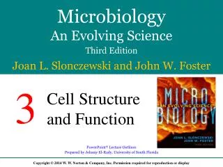
The Bacterial Cell: An Overview Cytoplasm = Consists of a gel-like network Cell membrane = Encloses the cytoplasm Cell wall = Covers the cell membrane Nucleoid = Non-membrane-bound area of the cytoplasm that contains the chromosome in the form of looped coils Flagellum = External helical filament whose rotary motor propels the cell
Biochemical Composition of Bacteria • All cells share common chemical components. • - Water • - Essential ions • - Small organic molecules • - Macromolecules • Cell composition varies with species, growth phase, and environmental conditions. Figure 1.1
The Cell Membrane Figure 3.7
Plasma membrane • Phospholipidbilayer: Contain a hydrophilic head and hydrophobic tail • The hydrophobic head is composed of glycerol and phosphate or other polar molecule • Tail contains fatty acid or hydrocarbon chains of differing chain length and chemistry
Membrane Constituents • Membranes have approximately equal parts of phospholipids and proteins. • A phospholipid consists of glycerol with ester links to two fatty acids and a phosphoryl head group. • - May have side chain Figure 3.8
Plasma membrane is a selective permeable barrier allows passage of some molecules into the cell and not all
Membrane Constituents Membrane proteins serve numerous functions, including: Structural support Detection of environmental signals Secretion of virulence factors and communication signals Ions and nutrient uptake proteins Proteins for ATP synthesis Photosythetic pigments, etc
Transport across the Cell Membrane Selective transport is essential for survival. - Small uncharged molecules, such as O2 and CO2, easily permeate the membrane by diffusion. - Water tends to diffuse across the membrane in a process called osmosis.
Polar molecules and charged molecules require transport through specific protein transporters. Figure 3.10 - Passive transport = Molecules move along their concentration gradient - Active transport = Molecules move against their concentration gradient - Requires energy
Transport across the Cell Membrane • Osmosis: Movement of water across the membrane • Simple diffusion: Movement of a solute from an area of high concentration to an area of low concentration. • Facilitative diffusion: Solute combines with a transporter protein in the membrane.
Membrane Transport • Passive diffusion (water, ions, O2, CO2) • Facilitated diffusion (protein carrier) • Active transporters (requires energy) • Group translocation (requires energy)
Movement across membranes Figure 4.17
Movement across membranes • Active transport of substances requires a transporter protein and ATP. • Group translocation of substances requires a transporter protein and PEP (phosphoenol pyruvate) as an energy source.
Membrane Lipids Phospholipids vary with respect to their phosphoryl head groups & their fatty acid side chains. Figure 3.11 Figure 3.12
Membranes also include planar molecules that fill gaps between fatty acid chains. In eukaryotic membranes, the reinforcing agents are sterols, such as cholesterol. In bacteria, the same function is filled by hopanoids, or hopanes. Figure 3.13
Archaea have the most extreme variations in phospholipid side-chain structures. - Ether links between glycerol and fatty acids - Hydrocarbon chains are branched terpenoids. Figure 3.14
The Cell Wall The cell wall confers shape and rigidity to the cell, and helps it withstand turgor pressure. The bacterial cell wall, or the sacculus, consists of a single interlinked molecule. Figure 3.16
The Cell Wall • Resists turgor pressure • Provides shape and rigidity
N-acetyl glucosamine (G) • N-acetyl muramic acid (M) • β-1,4 glycosidic • Peptide cross-link
Peptidoglycan Structure Most bacterial cell walls are made up of peptidoglycan (or murein). The molecule consists of: - Long polymers of two disaccharides called N-acetylglucosamine and N-acetylmuramic acid - The peptides form cross-bridges connecting the parallel glycan strands.
Most bacteria have additional envelope layers that provide structural support and protection. Envelope composition defines: - Gram-positive bacteria (thick cell wall) - Example: The phylum Firmicutes - Gram-negative bacteria (thin cell wall) - Example: The phylum Proteobacteria Gram-Positive and Gram-Negative Bacteria Figure 1.1
Capsule (not all species) - Made of polysaccharides S-Layer (not all species) - Made of protein or glycoproteins Thick cell wall - Amino acid cross-links in peptidoglycan - Teichoic acids for strength Plasma membrane Gram-Positive Cell Envelope Figure 3.18a
Gram - Positive cell walls • Teichoic acids: • Lipoteichoic acid links to plasma membrane • Wall teichoic acid links to peptidoglycan • May regulate movement of cations Figure 4.13b
Mycobacterial Cell Envelopes Mycobacterium tuberculosis and M. leprae have very complex cell envelopes. - Include unusual membrane lipids (mycolic acids) and unusual sugars (arabinogalactans) Figure 3.21
Gram-Negative Outer Membrane The thin peptidoglycan layer consists of one or two sheets. -Covered by an outer membrane, which confers defensive abilities and toxigenic properties on many pathogens Figure 3.18b
Eukaryotic Microbes Eukaryotic microbes possess their own structures to avoid osmotic shock. - Algae form cell walls of cellulose. - Fungi form cell walls of chitin. - Diatoms form exoskeletons of silicate. - Paramecia possess a contractile vacuole to pump water out of the cell.
Eukaryotic Microbes Figure 3.25
Gram positive vs Gram negative • Single or a thin layer of peptidoglycan • Outer membrane present • Teichoic acid absent • Lipopolysaccharide present. • Porins present • Thick peptidoglycan & suseptible to penicillin • No outer membrane present. • Teichoic acid present • No lipopolysaccharide present. • Porins absent