Download
1 / 50
550 likes | 822 Vues
New Detectors for PET Imaging of Small, Awake Animals. RatCAP Non-invasive wrist monitor Beta microprobe. Craig Woody Instrumentation Seminar March 12, 2003. Positron Emission Tomography. e +. e -. Radioisotope. Isotopes emit positrons with energies of a few hundred keV. e +.

E N D
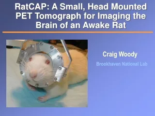
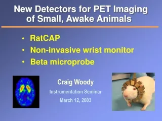
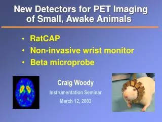
New Detectors for PET Imagingof Small, Awake Animals RatCAP Non-invasive wrist monitor Beta microprobe Craig Woody Instrumentation Seminar March 12, 2003
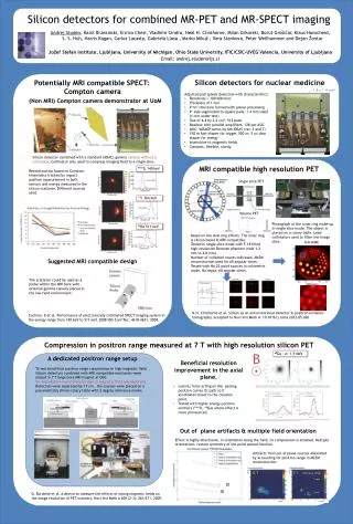
Positron Emission Tomography e+ e- Radioisotope Isotopes emit positrons with energies of a few hundred keV e+ range ~ few mm g g PET detects coincident 511 keV gamma rays PET Scanner
Positron Emitting Radiotracers Organic molecules are labeled with positron emitting isotopes and used as tracers Tracers can be used to study neurotransmitter activity in the brain
Neurotransmitter Activity in the Brain MAO A DA DA 11C-Cocaine DA DA DA DA DA DA DA DA DA DA DA DA DA DA DA DA DA DA signal Drugs like cocaine can block the re-uptake sites for neurotransmitters like dopamine which upsets the normal equilibrium and can cause effects of addiction
The Problem One wants to use PET to study the neurophysiological activity and behavior in laboratory animals in order to understand and treat these effects in humans. However, animals currently need to be anesthetized during PET imaging. • Cannot study animal behavior while under anesthesia • Anesthesia can greatly depress brain functions and • affect the neurochemistry that one is trying to study
Effects of Anesthesia H.Hideo et.al., Synapse 42 (2001) 273-280 D.B. Stout et al, UCLA 1998 Reduction in glucose metabolism with isoflurane in humans Similar effects are seen in the rat The effect of anesthesia on the uptake of β-CFT on dopamine transporters in the monkey brain.
RatCAP: RatConcious Animal PET A septa-less, full-ring tomograph with a diameter of 4 cm and an axial extent of 2 cm, suspended by a tether, which would allow nearly free movement of the awake animal Mockup of the portable ring on the head of a rat
The Collaboration Part of a larger project for Imaging The Awake Animal also involving motion tracking in both PET and MRI
The People Physics Craig Woody Sean Stoll Bill Lenz Mike Lenz Instrumentation Veljko Radeka Paul O’Connor Jean-Francois Pratte Bo Yu SUNY Stony Brook Sepideh Shokouhi Azael Villanueva Aarti Kriplani Chemistry David Schlyer Richard Ferrieri Mike Schueller Joanna Fowler Medical Paul Vaska Nora Volkow
Design Requirements • The tomograph ring must be light enough to be supported by the rat and allow reasonable freedom of movement • Light weight detectors (~ 125 g total weight) • Light weight electronics with low power dissipation New custom ASIC • High data rates and large singles background • Small field of view and large parallax effects • Limited sampling due to space and weight requirements • Must be rugged enough withstand activity of the rat
Support Structure Similar support structures are used in microdialysis experiments “Ratturn Bowl” Prototype support tether
Tomograph Ring Ring containing 12 block detectors Up to two layers of 5 mm deep crystals with APDs and integrated readout electronics 2x2 mm2 LSO crystals read out with APD arrays
Sensitivity Relative Scanner Sensitivities 25 Concorde rat brain UCLA region 20 BNL nanoPET Axial Acceptance Angle 15 (deg) 10 5 0 -4 -2 0 2 4 Comparison of sensitivity with other small animal PET scanners BNL RatCAP 4 cm Ø x 2 cm UCLA mPET 17 cm Ø x 2 cm centimeters • Intrinsic detector sensitivities are • essentially the same • Coincidence sensitivities are • proportional to axial acceptance Concord mPET R4 15 cm Ø x 8 cm P.Vaska
Parallax Effects 2 cm Due to the small diameter of the ring, the parallax error is a major factor in determining the spatial resolution
Spatial Resolution and Field of View P.Vaska Resolution at the edge of the rat brain Single layer - 10 mm ~ 2.5 mm Double layer - 5 mm ~ 1.9 mm Radial Spatial Resolution FWHM (mm) First prototype will be a single layer of 5 mm crystals Will sacrifice sensitivity for improved spatial resolution
Inter-Crystal Scatter and Cross Talk • Energy information in each crystal could be used to recover Compton events and correct for cross-talk between pixels • Collected data with ADC information and compared energy, position, time resolution and sensitivity : • DE (FWHM) : 21% 19% • time & position resolution ~ same • ~25% increase in coincidence sensitivity Conclusion: The slight improvement in resolution and increased sensitivity does not justify the added complexity in the ASIC P.Vaska et.al., 2002 MIC, to be published in IEEE TNS
Light Output and Energy Resolution Proteus unbonded 3M reflector CTI white powder reflector Studying different types of crystal arrays to optimize light output and energy resolution Proteus glued 3M reflector
Contributions to Energy Resolution 137Cs 662 keV g s/m ~ 7.5% Nsc = Number of scintillation photons produced Ncol = Number of photons collected Ne-h= Number of electron-hole pairs produced in APD dnoise = Dark current noise S.Stoll
Monte Carlo Study of Light Collection OPTICAD Program Maximum light collection near ends of crystal Very dependent on geometry and reflector properties S.Shokouhi
Comparison of Light Output and Energy Resolution of Different Crystals Average Light Output and Resolution of 4x8 LSO arrays FWHM 3M Reflector 3M Reflector S.Stoll Conclusion: Crystals wrapped with 3M reflector and air gap give the highest light output and best energy resolution
Avalanche Photodiodes Hamamatsu S8550 Improvements in Gain and Dark Current Jan ‘01 Jan ‘02 • Typical G ~ 50 • Npe ~ 1200 • ~ 60K signal electrons Expected noise in final ASIC ~ 500-600 e’s 4x8 array 1.6 x 1.6 mm2 active pixel area CT~ 10 pF
Gain and Quantum Efficiency Variation Measured with N2 laser + optical fiber Channel to channel gain variation s/m ~ 3.6 % Quantum efficiency variation s/m ~ 6.8 % Channel to channel differences dominated by quantum efficiency variation s/m ~ 6.4 % Gain + QE variation S.Stoll
Timing Resolution Need good time resolution (~ few ns) for coincidence timing to reject singles background 1 ns t ~ 70 ns S.Stoll B.Yu DT~ 5 ns fwhm Expect time resolution to improve considerably with new ASIC Presently dominated by noise in electronics setup
Test Setup with Crystal and APD Arrays Challenge is to put all of this on a chip ! Lab 2-93 in Physics Two arrays of 4x8 APDs and crystals using hybrid preamps and shapers and CAMAC DAQ system
RotatableStage for Taking Tomographic Data Source phantoms mounted on rotatable stage to simulate full tomographic ring
Tomographic Images 2 mm Measured with a ~2 mm diameter 68Ge point source and two gamma coincidence 52 samples (6.9 degrees) S.Shokouhi Spatial resolution is 2.1 mm FWHM
Comparison with MicroPET MicroPET resolution measured with the same ~ 2 mm point source Spatial Resolution MicroPET ~ 2.7 mm RatCAP ~ 2.1 mm P.Vaska & S.Shokouhi
Modeled Reconstructed Images Reconstruction Simulations using SimSet Image reconstructed using incomplete data set Image reconstructed using incomplete data set with interpolation Fully sampled image of four circular point sources S.Shokouhi
Electronics & Readout System • Tomograph ring with detectors and front end readout electronics are mounted to the head of the rat • Tether carries discriminator pulses, encoded addresses, high and low voltage power to a Data Collection Module which adds time stamp information
Tomograph Ring with Readout Electronics LSO blocks, APDs and front end readout electronics will be mounted on a PCB / Multi Chip Module with interconnecting flexstrip cable A.Kandasamy
So you want me to put my head in here ?…. A.Kandasamy
No analog information Single ZCD per channel Serial transmission from on-block electronics to a Data Collection Module (DCM) at the top of the tether DCM adds time stamp to each event and sends address and time information to a remote coincidence processor Individual links from each block to DCM Electronics + Readout System DCM P.O’Connor ~1.5 watts total power on ring
Custom Front End Readout Chip Custom ASIC in 0.18 μm CMOS gives ~ 4 mW per channel (~125 mW per chip) • Discriminator pulse is • encoded to give 5 bit • address • Leading edge of encoded • serial pulse train gives • time information J-F. Pratte P.O’Connor
First test chip delivered Feb ‘03 and ready for testing 2nd version submitted Nov ‘02 3rd version to be submitted March ‘03 Final version to be submitted fall ‘03 ASIC Production J.-F. Pratte Preamp circuit is contained within the yellow circle Final size will be ~ 4.3 x 1.6 mm
Alternative Approach: Light-sharing LSO/APD LSO slab APDs P.Vaska • LSO slab ~ 6 cm Ø, 10mm thick obtained from CTI • Large Area APDs Requires low-noise • Suppliers • Adv Phot, P-E, RMD • 3 RMD 8x8mm now in hand • Currently: setting up to measure energy & transverse spatial resolution The depth of interaction is determined directly without pixelating the crystals
Non-Invasive Wrist Monitor • A wide range of quantitative PET studies using tracer • kinetic modeling demand accurately measured • radiotracer concentration in arterial blood as a function • of time after injection, known as the Arterial Input Function. • The common method of measuring the input function is • the invasive withdrawal of blood from a wrist artery. • However, because of its health risks for both patients and • hospital personnel, it is not compatible with clinical studies. • A small ring tomograph similar to the RatCAP can be • used to image the artery and measure the input function
Wrist Anatomy For human studies, the input function will be taken from the radial artery in the wrist. Activity in the surrounding veins produce a significant background which can be rejected using the good spatial resolution of the wrist monitor. Phantom based on MRI images of the wrist with anatomically correct placement of the vessels A.Villanueva
Planar Images S.Shokouhi, 2002 MIC, submitted to IEEE TNS Measured with a 1 mm diameter 68Ge line source and two gamma coincidence 1 x 1 mm2 pixels
Simulated Input Function Measurement Measure activity as an aqueous solution of a 11C - labeled radioisotope passes between the two block detectors w/scatter w/o scatter Expected Input Function resolution for Wrist Monitor A.Villanueva
Beta Scintillation Microprobe • Radioisotopes used in PET emit positrons with energies • of a few hundred keV which have a range of several mm • in blood or tissue. This range is comparable to the spatial • resolution obtained in most PET cameras. • These positrons can be detected directly using plastic or • crystal scintillators. With crystal scintillators which have • high density, the positrons will range out, depositing all of • their energy in the crystal and producing a large signal. • Small scintillation probes can be used to directly measure • the radiotracer concentration in the blood or tissue. C.L.Woody et.al., IEEE Trans. Nucl. Sci NS-49 (2002) 2208-2212.
Comparison of LSO vs Plastic Scintillator Range and energy loss of positrons in LSO and plastic scintillator Response of LSO and plastic scintillation probes to betas (32P) and gamma rays (137Cs).
Sensitivity and Position Resolution Material LSO LSO Plastic Size 0.3 x 0.5 mm 0.5 x 1.5 mm 1.0x1.0 mm Volume 0.035 mm3 0.295 mm3 0.795 mm3 Sensitivity 10.1 20.7 51.2 (Hz/mCi/cc) Region of sensitivity around the probe can be selected by adjusting the readout threshold to improve spatial resolution
Probe Construction LSO microprobe consisting of 0.5 mm diameter x 1.5 mm long LSO crystal wrapped with several layers of white reflecting teflon and covered with polyester shrink tubing.
Rat Brain Studies with Microprobe Uptake of 11C-methylphenidate in the nucleus accumbens region of a rat brain with LSO probe nucleus accumbens ~ 2 mm
Input Function Measured with Microprobe Input function LSO microprobe (0.3 mm dia. x 0.5 mm) inserted inside an 18 gauge syringe needle for blood flow study. Activity of 11C-tyrosine measured in baboon blood flow during a PET scan using a syringe mounted LSO microprobe.
Summary • New detectors are needed for PET studies of the neurological behavior in live, awake animals • The RatCap will provide a new tool for carrying out these studies in laboratory rats. • Similar small ring tomographs can be used to measure the arterial input function in larger animals and humans • Small scintillation microprobes can be used to directly measure the positron activity in blood and tissue in live, awake animals with a spatial resolution comparable to or better than current PET tomographs
Neurochemistry 101 MAO A DA DA 11C-Cocaine DA DA DA DA DA DA DA DA DA DA DA DA DA DA DA DA MAO B DA DA signal MonoAmineOxydase-B (outside neuron) Deprenyl given to increase MOA-B Vesicules containing dopamine MonoAmineOxydase-A (inside neuron) Both MOA- A,B consume dopamine Dopamine transporters (re-uptake sites) (Cocaine, MP) Methylphenadate (MP) = Ritalin Ceretonin (neurrotransmitter) Present in frontal corrtex (Cerebellum) Dopamine receptors (raclopride) Only few % receptor sites occupied Striata High Dopamine concentration Raclopride concentrates at D2 receptor sites