Cardiovascular Disease
Cardiovascular Disease. 附一儿科 林洁英. General Consideration. Fetal Circulation. Change of Circulation after Birth. Cardiovascular Disease Congenital Heart Disease (CHD). Etiology of CHD. Diagnosis of CHD. Classification of CHD. Several Common CHD. Complication of CHD.

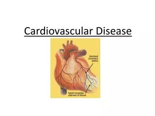
Cardiovascular Disease
E N D
Presentation Transcript
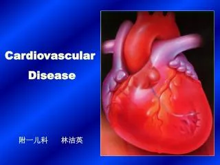
Cardiovascular Disease 附一儿科 林洁英
General Consideration Fetal Circulation Change of Circulation after Birth Cardiovascular Disease Congenital Heart Disease (CHD) Etiology of CHD Diagnosis of CHD Classification of CHD Several Common CHD Complication of CHD Treatment of CHD
胚胎时期的心脏发育 • 1.原始心脏于胚胎第2周开始形成,约于第4周起有循环 作用,至第8周房室隔已完全形成,即为四腔心。 • 房间隔形成(卵圆孔的存在;异常时形成房间隔缺损) • 室间隔形成(异常时形成室间隔缺损) • 大动脉形成(异常时形成可形成大动脉转位,主动脉骑跨等) • 2.心脏胚胎发育的关键时期:妊娠第2~8周。这一时期就是先天性心脏病形成的主要时期。
Fetal Circulation Diagram for normal fetal circulation 1. Left Atrium, LA 2. Left Ventricle, LV 3. Right Atrium, RA 4. Right Ventricle, RV 5. Superior Vena Cava, SVC 6. Inferior Vena Cava, IVC 7. Aorta, AO 8. Pulmonary Artery, PA
Fetal Circulation characters:(5 points) 1. Placenta: exchange of nutrition & oxygen 2. The resistance of pulmonary circulation> systemic circulation 3. Unclosed oval foramen, opened patent ductus 4. mixed blood inside the heart 5. nutrient organs,liver—heart—brain—body— upper extremities—lower extremities
The change of circulation after birth character:(4points) 1. Resistance of pulmonary circulation , resistance of systemic circulation 2. Close of oval foramen 3. Close of patent ductus 4. Independent systemic & pulmonary circulation are formed, no mixed blood inside the heart.
Endogenous factors Etiology of CHD External factors
(1). Endogenous factors Relate to heredity and chromosome (2). External factors Intrauterine infection
radioactive rays, drugs Prevent from virus infection at 1st trimester is important, because that the fetal heart developed 2~8 week after pregnancy.
Diagnosis of CHD 1 history virus infection radioactive rays medication 1.1 maternal pregnant history 1.2 common symptoms slight case serious case 1.3 age of onset Heart disease attack before 3 y--CHD Heart disease attack after 3y--RHD
2 Physical Examination 2.1 general manifestation 2.2 heart examination 2.3 peripheral vessel sign
3 Special Examination 3.1 X-ray examination 3.2 ECG 3.3 UCG
3. UCG Demonstrate the heart structure: Normal infantile M-mode UCG
3.4cardiac catheterization 3.5 cardioangiography 3.6 radionuclear angiography 3.7 Magnetic Resonance Imaging MRI
先天性心脏病的分类 左向右分流型 (潜伏青紫型) Classification of CHD 右向左分流型 (青紫型) 根据血循环的改变分三大类 无分流型 (无青紫型)
左向右分流(潜伏青紫型) left to right shunt(potential cyanosis type) 常见疾病:ASD、VSD、PDA。正常情况下,体循环压力>肺循环压力,血液从左向右分流而不出现紫绀,当剧哭、屏气或病理情况下,肺动脉或右室压力高于左心压力时,产生右向左分流而出现暂时青紫,故也称潜伏青紫型。体循环血流量减少,肺循环血流量增加,随着病情进展,可出现动力性肺高压,如肺循环阻力进行性增加,最终出现梗阻性肺高压,临床出现青紫,称艾森曼格(Eisenmenge)综合征。
右向左分流(青紫型) right to left shunt(cyanosis type) 常见疾病:法洛四联征、大动脉转位等。 由于畸形的存在,血液从右向左分流或大血管起源异常,使大量静脉血流入体循环。临床上出现持续性紫绀,故也称青紫型。
无分流型(无青紫型) no shunt(no-cyanosis type) 常见疾病:肺动脉辧狭窄、主动脉缩窄。 是指心脏的左、右两侧或动、静脉之间无异常分流
临床表现 1.左向右分流型:轻症患儿可无症状或症状轻微。常见表现生长发育迟缓,气促,活动耐力差。小婴儿则表现喂养困难,多汗、脸色苍白等,易患肺炎,心力衰竭等并发症。 2.右向左分流型:紫绀是最常见症状,其他有活动后气促、喜蹲踞、红细胞代偿性增多、杵状指等表现。易出现栓塞等并发症。
先天心脏病并发症 1.左向右分流:支气管肺炎,充血性心力衰竭,肺水肿,亚急性细菌性心内膜炎等。 2.右向左分流:脑脓肿,脑血栓,亚急性细菌性心内膜炎等。
先天性心脏病治疗原则 1.内科治疗:增强患儿体质,预防和减少感染的发生,防治并发症。 肺炎、心内膜炎要及早诊断,积极控制感染;心功能不全时强心、利尿、扩管等。 2.手术治疗:外科手术,介入治疗。
Several Common CHD 1Ventricular Septal Defect (VSD) 2 Atrial Septal Defect (ASD) 3 Patent Ductus Arteriosus (PDA) 4 Tetralogy of Fallot (TOF)
Ventricular Septal Defect most common CHD, in our country -- 50% 1、defect position 2、pathophysiology 3、clinical manifestation 4、X --ray examination 5、ECG 6、echocardiogram 7、cardiac catheterization 8、cardioangiography
1、Defect position depend on the defect position, type VSD as follows: (1)superior to the crista supraventricularis and inferior to aortic valve or pulmonary valve; (2)inferior to the crista supraventricularis; (3)posterior to tricuspid valve; (4)at muscular septum, may complicate several defect. (2)(3) are so called membranous VSD.
1. RV 2. LV 3. RA 4. LA 5. AO 6. PA 7. VSD The Anatomic classification of VSD (1st one)
2.Pathophysiology 1.左→右分流,肺循环量↑ 2.血流动力学改变与缺损大小及肺血管床状况有关。 小VSD(<0.5cm):分流量小,血流动力学变化不大; 中VSD(0.5~1.5cm):明显左→右,Qp>2~3倍Qs; 大VSD( > 1.5cm):分流量很大, Qp>3~5倍Qs 3.病情进展,肺循环量持续↑,动力性PH →梗阻性PH,右→左分流,艾森曼格综合征。
Pathophysiology A before pulmonary hypertension RA LA hypertrophy shunt PA RV LV hypertrophy (dilation) (blood volume )(ejection volume ) pulmonary RV systemic circulation (expansion) circulation (congestion) (insufficient blood supply) The hymodynamics of large VSD (1st one)
B After pulmonary hypertension RA LA shunt PA RV LV dilation expansion (hypertrophy) dynamic PA (mixed blood) hypertension systemic circulation organic PH The hymodynamics of large VSD (2nd one)
3、Clinical manifestation:owe to the defect size symptoms Small VSD -- defect < 0.5 cm, may no symptoms, good growth Middle VSD --defect 0.5 ~1.5 cm,large shunt volume, loss of systemic blood volume, interfere the grow & develoment. Large VSD -- defect > 1.5 cm, pulmonary hypertension left to right shunt right to left shunt cyanosis Eisenmenger’s syndrome
Signs: Small VSD: SM(L3—4) Middle -Large VSD: inspection: palpation:L3—4 ST percussion: auscultation:L3—4 Sm, P2 pulmonary hypertension Naturally close of VSD:20—50% of VSD may be closed before 5 y old.
4、X--ray Examination Middle--sized VSD (d = 0.9 cm)
X--ray Examination Large--sized VSD (2.2x2.5 cm)
X--ray Examination Large--sized VSD with pulmonary hypertension & heart failure( 2.5cm) Pulmonary pressure--14.4/10.4 kPa(108/78 mmHg)
6、UCG Parasternal long--axis echocardiographic view
UCG Apical four--chambers view
UCG Left to right shunted colored flow diagram
室缺的治疗 1.内科治疗:强调定期门诊随访,把握手术时机。心衰时,强心(洋地黄)、利尿、扩管(ACEI)等。 2.手术治疗:外科手术、介入手术。 手术指征:应尽早手术治疗:反复肺部感染、难以控制的心力衰竭、2 岁内肺动脉压力持续增高。 其余病例宜在学龄前手术治疗。 无症状小VSD可长期随访。
Several Common CHD 1Ventricular Septal Defect (VSD) 2Atrial Septal Defect (ASD) 3 Patent Ductus Arteriosus (PDA) 4 Tetralogy of Fallot (TOF)
Atrial Septal Defect (ASD) Account for 20 ~ 30 % of CHD,more females. 1. defect type 2. pathophysiology 3. clinical manifestation 4. X-ray examination 5. ECG 6. echocardiogram 7. cardioangiography 8. cardiac catheterization
1. defect type 分原发孔型房缺、继发孔型房缺、静脉窦型房缺三型。以继发孔型房缺最为常见,约占75%。缺损位于房间隔中心卵圆窝部位,也称中央型房缺。原发孔缺损常伴二尖瓣裂。
病理生理 1.分流量大小与缺损大小、两侧心房压力差及心室顺应性有关。 2.左→右分流,右心血流量↑,舒张期负荷↑,RA、RV增大,肺循环血量↑,压力↑,晚期可致梗阻性肺高压,出现艾森门格综合征。
pathophysiology SVC, IVC P V RA shunt via ASD LA (blood volume) blood volume RV (expansion) LV(blood volume PA(dilation) AO blood volume pulmonary congestion insufficient systemic blood supply Hemodynamics of ASD