Download

1 / 20
210 likes | 513 Vues
Trauma – Spinal Cord Injury. Sameer D. Khatri, MD. Learning Objectives. Correctly perform primary/secondary surveys and recognize physical signs of spinal cord injury Be aware of risk factors and understand how to manage spinal cord injuries Recognize and treat neurogenic shock

E N D
Trauma – Spinal Cord Injury Sameer D. Khatri, MD
Learning Objectives Correctly perform primary/secondary surveys and recognize physical signs of spinal cord injury Be aware of risk factors and understand how to manage spinal cord injuries Recognize and treat neurogenic shock Understand clinical decision rules to obtain radiographs for c-spine injuries
Introduction Spinal cord injury remains a devastating, epidemic event in modern society, despite early recognition, and treatment Injury usually leads to severe and permanent disability TSCI effects young adults disproportionately and is costly to society
Major Causes of TSCI in the U.S. Motor vehicle crashes: 47% Falls: 23% Violent acts like gunshot wounds: 14% Sports related accidents: 9% Other: 7%
Risk factors for TSCI Cervical spondylosis Atlantoaxial instability Congenital conditions like tethered cord Osteoporosis Spinal arthropathies like ankylosingspondylitis or rheumatoid arthritis
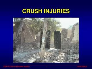
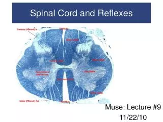
Pathophysiology • Injury to the bony vertebral column due to: • Fracture • Dislocation • Ligamentous tears • Herniation or disruption of intervertebral disc
Initial Evaluation and Treatment • Stabilize using ABCD prioritization • Assume spine injury if: • Head injury present • Unconscious or confused • Spinal cord pain present • Extremity weakness is present • Loss of sensation • Use log-roll, backboard and rigid c-collar
Spinal and Neurogenic Shock • Transient loss of spinal cord function can follow TSCI • Assume hypotension due to hemorrhage • Consider neurogenic shock if: • Hypotension not responding to fluids and/or bradycardia • Stabilizing BP may require pressors • Stabilizing HR may require atropine or pacing
Emergency Management • Continue prioritizing care using ABCDs • Continuous vital sign monitoring • Be aware that 1/3 of cervical injuries require intubation within 24 hours (Gardner, 1986) • Address hypoxia • Address hypotension • Complete neurologic exam ASAP • Asses for bladder distension and insert urinary catheter ASAP
Imaging • Plain films can be used to assess C-spine • AP, lateral, and odontoid views (minimum) • NEXUS Low-risk Criteria or Canadian C-spine rule –> imaging needed? • CT usually done if C-spine plain films inadequate or for lower spine injuries
NEXUS Low Risk Criteria • C-spine imaging not necessary if: • Absence of posterior midline cervical tenderness • Normal level of alertness • No evidence of intoxication • No abnormal neurologic findings • No painful distracting injuries • Altered LOC defined as: • Glasgow coma scale score below 15 • Disorientation to person, place, time, or events • Inability to remember three objects at 5 minutes • Delayed or inappropriate response to external stimuli
Canadian C-spine Rule • Condition One – Perform imaging if: • Age 65 years or older • Dangerous mechanism of injury • Parasthesias in the extremities • Condition Two – Assess low risk factors: • Simple rear end MVA • Sitting position in ED • Ambulatory at any time • Delayed onset of neck pain • Absence of midline cervical spine tenderness • Condition Three • Test active range of motion. If not able to rotate neck 45 degrees L&R. If able to do so, don’t need imaging.
Treatment • Immediate surgical consultation is recommended for all cases of TSCI • Intensive medical care and continuous monitoring required in the ICU setting to minimize complications / disability • Insufficient evidence to administer steroids
Summary • Injury to the spinal cord should be assumed in any trauma situation • Precautions using C-collars, backboards, and log-rolling is advised to prevent worsening injury • C-spine can be ruled out with adequate plain films and avoided altogether in certain using clinical decision rules • ABCDs scheme should be used to prioritize care
References • Hansebout, Robert R., Kachur, Edward Acute traumatic spinal cord injury. UpToDate, Waltham, MA, 2012. • Gardner BP, Watt JW, Krishnan KR. The artificial ventilation of acute spinal cord damaged patients: a retrospective study of forty-four patients. Paraplegia 1986; 24:208 • American College of Surgeons, ATLS Student Guide, 9th Edition, Sep 2012.
Simulation Training Assessment Tool (STAT)– Penetrating Neck Injury Sameer D. Khatri, MD
Simulation Training Assessment Tool (STAT) – Spinal Cord Trauma Learning Objectives: Recognize and appropriately manage spinal cord trauma Understand how to recognize and manage neurogenic shock Consult Neuro Surgery or Ortho Spine Surgery for disposition Date: Instructor(s): Learner(s):
Debriefing Notes • A transient loss of spinal cord function can occur following spinal column injury • Clinicians must assume that hypotension following trauma results from hemorrhage and treat as such • Neurogenic shock from SCI may cause hypotension and bradycardia • Use of pressors my be necessary to stabilize BP • Use of atropine or pacing may be needed for HR • Secondary survey: • Inspect entire body starting with head • Palpate entire spine and paraspinal musculature for tenderness, step-offs, or deformity • Perform a focused but systematic neuro exam to asses for symmetry in movements, priapism, abnormal breathing pattern, symmetry of peripheral strength, senasation, reflexes, sensory deficits, bowel, or bladder incontinence • Adequate plain xrays are essential to rule out c-spine injuries • AP, Lat, and odontoid views needed at minimum • CT scan required if visualization is inadequate • Use ABCs approach when reading plain film imaging • NEXUS Low-risk Criteria (NLC); No radiographs if • Absence of posterior midline cervical tenderness • Normal level of alertness • No evidence of intoxication • No abnormal neurologic findings • No painful distracting injuries • Altered LOC in NEXUS defined as: • Glasgow coma scale below 15 • Disorientation to person, place, time, or events • Inability to remember three objects at five minutes • Delayed or inappropriate response to external simuli • Canadian C-spine Rule: • Condition One – Perform imaging if: • Age 65 years or older • Dangerous mechanism of injury (falls, high speed MVA, axial load to head, ejection, collisions) • Parasthesias in the extremities • Condition Two – Assess low risk factors: • Simple rear end MVA • SAmbulatory at any time • itting position in ED • Delayed onset of neck pain • Absence of midline cervical spine tenderness • Condition Three • Test active range of motion. If not able to rotate neck 45 degrees L&R. If able to do so, don’t need imaging.
Additional Instructor Notes Case Synopsis • 25 yo male suffered a traumatic spinal cord injury after a ski related accident where he was found down. On arrival by first responders, pt was found to have severe upper back pain but was still alert and oriented X 3 with a GCS of 15. He was placed in C-collar and backboard and taken to the ED. En route, he was found to have transient hypotension and given a fluid bolus which rapidly normalizing his pressures. Learner must continue through the ABCDs and the rest of the critical actions. • Consider telling the learner that the patient was not moving his lower extremities bilaterally. Personnel and Roles • Instructor—Introduces case, switches to “EMS” as case begins, provides ancillary data as requested and plays “Surgeon” at the end of the case • Assistant (may be resident) —Acts voice of patient and manages monitor (tell Primary Learner that the assistant will be the patient’s voice prior to entry). Can also substitute an actor who can respond to the questions asked • Primary learner (resident)—Is the responding doctor. May lead the Trauma Team response or act as sole provider, depending on how your institution manages trauma • Secondary learners (residents)– Prompt primary learner to assign roles, e.g. Airway, Procedures, Nursing etc prior to beginning case. Props/Supply Checklist • Sim Man w/ C-collar on and strapped onto a backboard (can substitute for a live actor) • Moulage as needed to simulate trauma sustained while skiing and running into a tree • Airway equipment– NRB, laryngoscope, ETT 8.0, suction, BMV, RSI drugs • Consideration for making Sim Man have priapism Supporting Stimuli • EMS Run Sheet– give to learner at start of case • Point of care labs– give to learner only if ordered, after credible time lapse • Imaging for C-spine, T- and/or L-spine– give to learner only if ordered, after credible time lapse
EMS Sheet • 25 yo male found down while skiing the slopes • BP115/70 • HR90 • RR22 • POX98% on RA • GCS15 • Transient hypotension – responded with fluid bolus