Spinal cord injury
Spinal cord injury. HOANG TRONG AI QUOC, MD EMERGENCY DEPARTMENT HUE CENTRAL HOSPITAL, VIETNAM 2011. Objectives. Describe the basic spinal anatomy and physiology Evaluate a patient with suspected spinal injury Identify the common types of spinal injuries and their X-ray features.

Spinal cord injury
E N D
Presentation Transcript
Spinal cordinjury HOANG TRONG AI QUOC, MD EMERGENCY DEPARTMENT HUE CENTRAL HOSPITAL, VIETNAM 2011
Objectives • Describe the basic spinal anatomy and physiology • Evaluate a patient with suspected spinal injury • Identify the common types of spinal injuries and their X-ray features. • NEXUS and Canadian C-spine rules • Stable vs Unstable fractures • Appropriately manage the spinal-injured patient during the first hour from injury. • Determine the appropriate disposition of the patient with spine trauma
Introduction • Vertebral column injury, with or without neurological deficits, must always be sought and excluded in a patient with • Multiple trauma. • Any injury above the clavicle • Age and gender: • 65-80% occur in men • 60% occur in 16-30 years of age • Mechanisms: • MVC (48%) • Falls (21%) • Penetrating injuries (15%) • Sports injuries (14%)
Epidemiology • 40% of trauma patients with neuro deficits will have temporary or permanent SCI • Many more vertebral injuries that do not result in cord injury • 10-15% have non-contiguous injuries • Multiple injuries in non-adjacent vertebrae • Most commonly injured vertebrae • C5-C7 • C1-C2 • T12-L2
Beware • Excessive manipulation and inadequate immobilization of a patient with a spinal cord injury can cause additional neurological damage and worsen the patient’s outcome
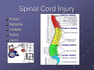
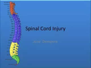
Anatomy and physiology • The spinal column: 7 cervical, 12 thoracic, and 5 lumbar vertebrae as well as the sacrum and the coccyx. • For many reasons, the cervical spine is most vulnerable to injury • The thoracolumbar junction is a fulcrum between the inflexible thoracic region and the stronger lumbar levels. This makes it more vulnerable to injury, with 15% of all spinal injuries occurring in this region.
Anatomy and physiology • Cervical Spine • 7 vertebrae • very flexible • C1: also known as the atlas • C2: also known as the axis
Anatomy and physiology • Thoracic Spine • 12 vertebrae • ribs connected to spine • provides rigid framework of thorax
Anatomy and physiology • Lumbar Spine • 5 vertebrae • largest vertebral bodies • carries most of the body’s weight • Sacrum • 5 fused vertebrae • common to spine and pelvis • Coccyx • 4 fused vertebrae • “tailbone”
Anatomy and physiology • Vertebral body • posterior portion forms part of vertebral foramen • increases in size from cervical to sacral • spinous process • transverse process • Vertebral foramen • opening for spinal cord • Intervertebral disk • shock absorber (fibrocartilage)
Anatomy and physiologySpinal ligament • Intrasegmental • - Ligamentum flavum • - Intertransverse ligament • - Interspinous ligament • Intersegmental • ALL • PLL • Supraspinous ligament
Anatomy and physiologySpinal Cord • Spinal cord ends at L1 • Three tracts can be readily assessed clinically. - The corticospinal tract - The spinothalamic tract - The posterior columns • Complete spinal cord injury: no sensory or motor function below a certain level, • Incomplete spinal cord injury: If any motor or sensory function remains, prognosis for recovery is much better. • Sparing of sensation in the perianal region (sacral sparing) may be the only sign of residual function.
Anatomy and physiologySpinal Cord • Blood supplied by vertebral and spinal arteries • Gray matter: core pattern resembling butterfly • White matter: longitudinal bundles of myelinated nerve fibers
Anatomy and physiologySpinal Cord • Thoracic and lumbar levels supply sympathetic nervous system fibers • Cervical and sacral levels supply parasympathetic nervous system fibers
Anatomy and physiologySpinal Cord Pathways • Spinothalmic Tracts (anterolateral) • Convey nerve impulse for sensing pain, temperature & light touch • Impulses cross over in the spinal cord not the brain • Lateral tracts :conduct impulses of pain and temperature • Anterior tracts: carry impulses of light touch and pressure
Anatomy and physiologySpinal Cord Pathways • Ascending Nerve Tracts (sensory input) • Carry sensory impulses from body structures • Posterior column (dorsal) • Conveys nerve impulses for proprioception, discriminative touch, pressure, vibration, & two-point discrimination • Cross over at the medulla from one side to the other (impulses from left side of body ascend to the right side of the brain)
Anatomy and physiologySpinal Cord Pathways • Descending Motor Tracts (motor output) • Conveys motor impulses from brain to the body • Pyramidal tracts: Corticospinal & Corticobulbar • Corticospinal tracts • cause precise voluntary movement and skeletal muscle activity • lateral tract crosses over at medulla - Extrapyramidal tracts Rubrospinal, pontine reticulospinal, medullary reticulospinal, lateral vestibulospinal and tectospinal
Example Motor and Sensory Pathways To thalamus and cerebral cortex (sensory) Spinothalmic tract Motor Cortex Brain Stem Posterior column Corticospinal tract Spinal Cord LMN Pain - Temp Proprioception (conscious) Example Motor Pathway (corticospinal tract)
Spinal Nerves • 31 pairs originate from the spinal cord • Carry both sensation and motor function • Named according to level of spine from where they arise • Cervical 1-8 • Thoracic 1-12 • Lumbar 1-5 • Sacral 1-5 • Coccygeal 1
Cord level • C2 – C7 = add +1 for cord level • T1 – T6 = add +2 • T7 – T9 = add +3 • T10 = L1, L2 level • T11 = L3, L4 level • L1 = sacro coccygeal segments
Dermatomes and key muscles • A dermatome is the area of skin innervated by the sensory axons within a particular segmental nerve root. They are important to determine level of injury
Dermatomes and key muscles • C5: Deltoids/biceps • C6: Wrist extensors • C7: Elbow extensors • C8: Finger flexors • T1: Finger Abductors • L2: Hip flexors • L3: Knee extensors • L4: Ankle dorsiflexors • L5: Long toe extensors • S1: Ankle plantar flexors
Common mechanism • Compression • Flexion • Extension • Rotation • Lateral bending • Distraction • Penetration
Suspect spinal injury with... • Sudden decelerations (MVCs, falls) • Compression injuries (diving, falls onto feet/buttocks) • Significant blunt trauma (football, hockey, snowboarding, jet skis) • Very violent mechanisms (explosions, cave-ins, lightning strike) • Unconscious patient • Neurological deficit • Spinal tenderness
High index of suspicion… • Missed or delayed diagnosis most often attributed to: • failure to suspect injury • inadequate radiology • incorrect interpretation of radiographs • A missed spinal injury can have devastating long term consequences • As such, spinal column injury must therefore be presumed until it is excluded
Spinal stabilization and management: pre hospital • Protect spine at all times during the management of patients with multiple injuries. • Up to 15% of spinal injuries have a second, possibly non adjacent, fracture elsewhere in the spine • Ideally, whole spine should be immobilized in neutral position on a firm surface. • Can be done manually or with a combination of semi-rigid cervical collar, side head supports, long spine board and strapping.
Spinal stabilization and management: pre hospital • PROTECTION => PRIORITY • DETECTION => SECONDARY • Rigid cervical collar • “Log-rolling” • Rigid transportation board • Rigid transfer slides
Spinal stabilization and management: pre hospital • Immobilization devices should not take precedence over life saving procedures • If neck is not in the neutral position, alignment should be made. • If the patient is awake and cooperative, actively move their neck into line themselves. • If the patient does not want to move neck because of pain – do not force movement of neck • Initial immobilization of C-spine with a hard-collar is a priority during extrication • Long spine boards are valuable primarily for extrication from vehicles. • Rapid evacuation to a trauma center
Spinal stabilization and management: hospital • PROTECTION => PRIORITY • DETECTION => SECONDARY • Rigid cervical collar • “Log-rolling” • Rigid transfer slides
Spinal stabilization and management: hospital • Spinal immobilization is a priority in trauma, spinal clearance is not • The spine should be assessed and cleared when appropriate, given the injury characteristics and physiological state • Imaging the spine does not take precedence over life saving diagnostic and therapeutic procedures
Clinical evaluation • Inspection and palpation: Occiput to Coccyx • Tenderness to the vertebrae • Gap or Step-off (both very rare) • Edema and bruising • Spasm of associated muscles • Neurological assessment • Sensation • Motor • Reflexes • Rectal examination
Assessment • Sensory: Dermatomes • Motor: Key muscles
AssessmentRectal tone • Tone: the presence of rectal tone in itself does not indicate an incomplete injury • Sensation • Volition: A voluntary contraction of the sphincter or the presence of rectal sensation presence of communication lower spinal cord- supraspinal centers favorable prognosis • Bulbocavernosus reflex: • Positive: presence of reflex lack of supraspinal input to the sacral outflow complete spinal injury • Negative: absent in spinal shock
SCI GRADING SYSTEMASIA: AMERICAN SPINAL INJURY ASSOCIATION • Neurological Classification: • Use the ASIA International standards • Motor and sensory assessment • ASIA Impairment Scale (A-E) • Clinical Syndromes (patterns of incomplete injury)
SCI GRADING SYSTEMASIA IMPAIRMENT SCALE • A = Complete: No motor or sensory function is preserved in the sacral segments S4-S5 • B = Incomplete: Sensory but not motor function is preserved below the neurological level and includes sacral segments S4-5 • C = Incomplete: Motor function is preserved below the neurological level, and more than half of key muscles below the neurological level have a muscle grade less than 3 • D = Incomplete: Motor function is preserved below the neurological level, and at least half of key muscles below the neurological level have a muscle grade of 3 or more • E = Normal: motor and sensory function are normal
SCI General Assessment • ABCs • Airway and/or Breathing impairment • Inability to maintain airway • Apnea • Diaphragmatic breathing • Cardiovascular impairment • Neurogenic Shock • Hypotension and bradycardia • Patient appears warm and dry • Hypoperfusion
SCI General Assessment • Neurologic Status: • Level of Consciousness • Brain injury also? • Cooperative • No impairment (drugs, alcohol) • Understands & Recalls events surrounding injury • No Distracting injuries • No difficulty in communication
SCI General Assessment • Assess Function & Sensation • Palpate over each spinous process • Sensation (Position and Pain) • weakness, numbness, paresthesia • pain (pinprick), sharp vs dull, symmetry • Motor function • Shrug shoulders • Spread fingers of both hands and keep apart with force • “Hitchhike” {T1} • Foot plantar flexors (gas pedal) {S1,2} • Priapism- Reflexes
Spinal Cord Injuries • Primary Injury • occurs at the time of injury • may result in • cord compression • direct cord injury • interruption in cord blood supply • Secondary Injury • occurs after initial injury • may result from • swelling/inflammation • ischemia • movement of body fragments
Spinal Cord Injuries • Cord concussion & Cord contusion • temporary loss of cord-mediated function • Cord compression • decompression required to minimize permanent injury (may have permanent injury) • Laceration • permanent injury dependent on degree of damage • Hemorrhage • may result in local ischemia
Spinal Cord Injuries • Cord transection • Complete • all tracts disrupted • cord mediated functions below transection are permanently lost • determined ~ 24 hours post injury • possible results • quadriplegia • paraplegia
Terminology • Paraplegia • loss of motor and/or sensory function in thoracic, lumbar or sacral segments of SC (arm function is spared) • Quadriplegia • loss of motor and/or sensory function in the cervical segments of SC
Spinal Cord Injuries • Cord transection • Incomplete • some tracts and cord mediated functions remain intact • potential for recovery of function • Possible syndromes • Brown-Sequard Syndrome • Anterior Cord Syndrome • Central Cord Syndrome
Brown Sequard Syndrome • Incomplete Cord Injury • Injury to one side of the cord (Hemisection) • Often due to penetrating injury or vertebral dislocation • Complete damage to all spinal tracts on affected side • Prognosis for recovery is variable
Brown Sequard Syndrome • Exam Findings • Ipsilateral loss of motor function motion, position, vibration, and light touch • Contralateral loss of sensation to pain and temperature • Bladder and bowel dysfunction (usually short term)
Anterior Cord Syndrome • Anterior Spinal Artery Syndrome • Supplies the anterior 2/3 of the spinal cord to the upper thoracic region • caused by bony fragments or pressure on spinal arteries
Anterior Cord Syndrome • Exam Findings • Variable loss of motor function and sensitivity to pinprick and temperature • loss of motor function and sensation to pain, temperature and light touch • Proprioception (position sense) and vibration are preserved
Central Cord Syndrome • Usually occurs with a hyperextension of the cervical region • Exam Findings • weakness or paresthesias in upper extremities but normal strength in lower extremities • varying degree of bladder dysfunction
Cauda Equina Syndrome • Injury to nerves within the spinal cord as they exit the lumbar and sacral regions • Usually fractures below L2 • Specific dysfunction depends on level of injury • Exam Findings • Flaccid-type paralysis of lower body • Bladder and bowel impairment