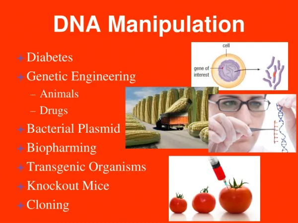
Gene Manipulation Concepts by Dr. G. Muralitharan - Assistant Professor
Learn about genetic engineering techniques, recombinant DNA technology, cloning, genomic DNA isolation, and nucleic acid preparation. Understand essential tools and methods for gene manipulation, DNA purification, and nucleic acid isolation. Discover the fundamental protocols and considerations for nucleic acid preparation.

Gene Manipulation Concepts by Dr. G. Muralitharan - Assistant Professor
E N D
Presentation Transcript
Gene Manipulation Concepts Dr. G. Muralitharan Assistant Professor Dept. of Microbiology Bharathidasan University Tiruchirappalli 620024 E-mail: drgm@bdu.ac.in
Genetic Engineering • Using in vitro techniques to alter genetic material in the laboratory • Basic techniques include • Restriction enzymes • Gel electrophoresis • Nucleic acid hybridization • Nucleic acid probes • Molecular cloning • Cloning vectors
Recombinant DNA Technology • Methods for isolating, manipulating, and amplifying identifiable DNA sequences. • Allows us to study the structure and function of individual genes. • Allows for the directed genetic manipulation of organism (modify gene function, insert novel genes)
Cloning • Clone: a collection of molecules or cells, all identical to an original molecule or cell • To "clone a gene" is to make many copies of it - for example, in a population of bacteria • Gene can be an exact copy of a natural gene • Gene can be an altered version of a natural gene • Recombinant DNA technology makes it possible • Allows for in vitro manipulation of a individual gene
Tools need for cloning • cDNA or genomic library (source of DNA to cut) • Plasmid (where you want to paste it) • Restriction enzymes (scissors) • DNA ligase (paste) • E. coli (biological machine needed to amplify DNA)
Plasmid DNA is the genetic material of most organisms (from bacteria to humans) Chromosome: Most bacteria have one circular DNA chromosome ranging in size from 1,000 to 8,000 kilobase pairs. Plasmid: Extrachromosomal genetic element also made of a circular DNA molecule. Bacterial Genome: The collection of all of the genes present on the bacteria’s chromosome or its extrachromosomal genetic elements.
Genomic DNA & Plasmid DNA isolation • Genomic DNA preparation overview • Plasmid DNA preparation • DNA purification • Phenol extraction • Ethanol precipitation
Nucleic Acid PreparationApplication? • DNA • Amplification methods (PCR, LCR) • Restriction enzyme digest • Hybridization methods (Southern analysis) • Sequencing
Nucleic Acid Preparation - Considerations • What is the size or volume of each sample? • Amount of DNA or RNA required • Equipment and tube sizes required • How many samples are being processed? • Capacity of the centrifuge • Isolation method speed • Is a high-throughput or automated system available? • 96-well plate methods • Walk-away or semi-automation
Nucleic Acid PreparationChoosing an Isolation Method • Important factors are: • Processing speed • Ease of use • Yield of DNA or RNA • Quality of DNA and RNA prepared (amplification performance) • Shelf life/storage conditions • Quality assurance criteria • Cost of preparation
Basic Protocol • Most DNA extraction protocols consist of two parts • A technique to lyse the cells gently and solubilize the DNA • Enzymatic or chemical methods to remove contaminating proteins, RNA, or macromolecules • In plants, the nucleus is protected within a nuclear membrane which is surrounded by a cell membrane and a cell wall. Four steps are used to remove and purify the DNA from the rest of the cell. • Lysis • Precipitation • Wash • Resuspension
DNA purification: overview cell harvest and lysis cell growth DNA concentration DNA purification
Bacterial genomic DNA prep: cell extract • Lysis: • Detergents • Organic solvent • Proteases (lysozyme) • Heat “cell extract”
Cell Lysis Buffer - action • Cell Lysis Buffer - Non-ionic detergent, Salt, Buffer, EDTA designed to lyse outer cell membrane of cells, but will not break down nuclear membrane. • EDTA (Ethylenediaminetetraacetic disodium salt) is a chelating agent of divalent cations such as Mg2+. Mg2+is a cofactor for Dnase nucleases. If the Mg2+is bound up by EDTA, nucleases are inactivated. • Proteinase K - used to remove most of the proteins. By digesting with proteolytic enzymes such as Pronase or proteinase K, which are active against a broad spectrum of native proteins, before extracting with organic solvents. Protienase K is approximately 10 fold more active on denatured protein. Proteins can be denatured by SDS or by heat.
Organic Extraction • Traditionally, phenol: chloroformis used to extract DNA. • When phenol is mixed with the cell lysate, two phases form. DNA partitions to the (upper) aqueous phase, denatured proteins partition to the (lower) organic phase. • Phenol:Denatures proteins and solubilizes denatured proteins
Organic Extraction Reagents • Phenol/Chlorform - The standard way to remove proteins from nucleic acids solutions is to extract once with phenol, once with a 1:1 mixture of phenol and chloroform, and once with chloroform. This procedure takes advantage of the fact that deproteinization is more efficient when two different organic solvents are used instead of one. • Also, the final extraction with chloroform removes any lingering traces of phenol from the nucleic acid preparation. • Phenol is highly corrosive and can cause severe burns.
Organic Extraction Reagents • Phenol - often means phenol equilibrated with buffer (such as TE) and containing 0.1% hydroxyquinoline and 0.2% b-mercaptoethanol (added as antioxidants. The hydoxquinoline also gives the phenol a yellow color,making it easier to identify the phases (layers). • Chloroform - often means a 24:1 (v/v) mixture of chloroform and isoamyl alcohol. The isoamyl alcohol is added to help prevent foaming. • The Phenol/Chloroform/Isoamyl Alcohol ratio is 25:24:1
Concentrating the genomic DNA 70% final conc. “spooling” Ethanol precipitation
Concentrating DNA – Alcohol Precipitation • The most widely used method for concentrating DNA is precipitation with ethanol. The precipitate of nucleic acid, forms in the presence of moderate concentrations of monovalent cations (Salt, such as Na+), is recovered by centrifugation and redissolved in an appropriate buffer such as TE. • The technique is rapid and is quantitative even with nanogram amounts of DNA. • The four critical variables are the purity of the DNA, its molecular weight, its concentration, and the speed at which it is pelleted. • DNA a concentrations as low as 20 ng/ml will form a precipitate that can be quantitatively recovered. • Typically 2 volumes of ice cold ethanol are added to precipitate the DNA.
Concentrating DNA – Alcohol Precipitation • Very short DNA molecules (<200 bp) are precipitated inefficiently by ethanol. • The optimum pelleting conditions depend on the DNA concentration. Relatively vigorous microcentrifuge steps such as 15 minutes at or below room temperature at 12,000 rpm are designed to minimized the loss of DNA from samples with yields in the range of a few micrograms or less. • Solutes that may be trapped in the precipitate may be removed by washing the DNA pellet with a solution of 70% ethanol. To make certain that no DNA is lost during washing, add 70% ethanol until the tube is 2/3 full. Vortex briefly, and recentrifuge. After the 70% ethanol wash, the pellet does not adhere tightly to the wall of thetube, so great care must be taken when removing the supernatant.
Concentrating DNA – Alcohol Precipitation • Isopropanol (1 volume) may be used in place of ethanol (2 volumes) to precipitate DNA. Precipitation with isopropanol has the advantage that the volume of liquid to be centrifuged is smaller. • Isopropanol is less volatile than ethanol and it is more difficult to remove the last traces; moreover, solutes such sodium chloride are more easily coprecipitated with DNA when isopropanol is used.
Separation by Salting Out Salting out method: • Celllysis. • Protein digestion by proteinase enzyme. • Protein precipitation by high salt concentration. • Centrifugationwill remove the precipitated proteins. • The supernatantcontains the DNA. • DNA is then precipitated by adding ethanol. • The precipitated DNA is resuspended in the desired buffer.
DNA Isolation MethodsSolid Phase Procedures • Uses solid support columns, magnetic beads, or chelating agents • Solid support columns: Fibrous or silica matrices bind DNA allowing separation from other contaminants. • Magnetic beads: DNA binds to beads; beads are separated from other contaminants with magnet. • Chelating resins • Advantages: • Fast and easy, no precipitation required
DNA purification: silica binding Binding occurs in presence of high salt concentration, and is disrupted by elution with water
What Does Qiagen silica Do? http://www.qiagen.com/resources/info/qiagen_purification_technologies_1.aspx Greenspoon, S. A., M. A. Scarpetta, M. L. Drayton, and S. A. Turek. 1998 . QIAamp spin columns as a method of DNA isolation for forensic casework. J Forensic Sci 43 (5): 1024–30.
Silica-Based Extraction Pro: • Quick • Highly purified DNA Con: • Multiple sample transfer • Increase risk of contamination
Magnetic Beads • Magnetic beads are coated with DNA antibodies to bind to DNA:
Magnetic Beads • Automated version:
Magnetic Beads Pro: • Very fast, may be automated • Highly purified DNA • Excellent for liquid blood Con: • Cannot be used directly on stain • i.e. need to remove cells from stain substrate (cloth, etc.) • Very expensive
Non-Organic DNA Extraction • Does not use organic reagents such as phenol or chloroform. • Digested proteins are removed by salting out with high concentrations of LiCl. • However, if salts are not adequately removed, problems could occur with the RFLP procedure due to alteration of DNA mobility (band shifting)
Non-Organic DNA Extraction Procedure • Cell Lysis Buffer - lyse cell membrane, nuclei are intact, pellet nuclei. • Resuspend nuclei in Protein Lysis Buffer containing a high concentration of Proteinase K. Lyse nuclear membrane and digest protein at 65oC for 2 hours. Temperature helps denature proteins, and Proteinase K auto digests itself • To remove proteinaceous material, LiCl is added to a final concentration of 2.5 M, and incubated on ice. Proteins precipitate out and are pelleted by centrifugation.
Non-Organic DNA Extraction Procedure 4. DNA remains in solution. Transfer supernatant to a new tube, care must be taken not to take any of protein pellet. 5. DNA is precipitated by the addition of room temperature isopropanol. LiCl will not precipitate with DNA. 6. Precipitated DNA is washed with 70% ethanol, dried under vacuum and resuspended in TE buffer.
Chelex Extraction Chelex 100, Molecular Biology Grade resin from BioRad is a highly pure, nuclease and ligase inhibitor-free chelating resin, certified not to interfere with downstream PCR. Specifically designed to complement the inherent requirements of PCR, this pure, pipettable, small-scale resin is ready for downstream use. Ensuring the complete removal of PCR inhibitors, contaminating metal ions that catalyze the digestion of DNA
Chelex Extraction • Chelex 100 is an ion exchange resin that is added as a 5% solution (wt/vol). • Chelex is composed of styrene divinylbenzene copolymers containing paired iminodiacetate ions that act as chelating groups in binding polyvalent metal ions such as magnesium (Mg2+). • By removing the Mg2+ from the reaction, nucleases are inactivated and the DNA is protected.
Chelex Extraction • Chelex 100 is an ion exchange resin that is added as a 5% solution (wt/vol). • Chelex is composed of styrene divinylbenzene copolymers containing paired iminodiacetate ions that act as chelating groups in binding polyvalent metal ions such as magnesium (Mg2+). • By removing the Mg2+ from the reaction, nucleases are inactivated and the DNA is protected.
Chelex Extraction • A 5% solution of Chelex is added to a blood stain or liquid blood and incubated at 56oC for 30 minutes. This step is used to lyse red cells and remove contaminants and inhibitors such as heme and other proteins. • The sample is then heated at 100oC for 8 minutes. This causes the DNA to be denatured as well as disrupting membranes and destroying cellular proteins. • The tube containing the Chelex is centrifuged, the resin is pelleted, the supernatant containing the DNA is removed.
Chelex Extraction • The Chelex extraction process denatures double stranded DNA and yields single stranded DNA, and thus cannot be used for the RFLP procedure. • It is advantageous for PCR-based typing methods because it removes inhibitors of PCR and can be done in a single tube, which reduces the potential for laboratory-induced contamination and sample switching. • Care should be taken not to have any residual Chelex with the DNA extract, since Mg2+ is required for the Taq Polymerase.
Chelex extraction Pros: • Relatively fast • Can extract directly from cloth (stain) • Minimizes contamination – uses only a single tube • Removes PCR inhibitors Con: • Results in single-stranded DNA – not useful for RFLP
Genomic DNA prep in plants -- how get rid of carbohydrates? CTAB: Cationic detergent (MC 6.61-6.62) CH3 CH3 (low ionic conditions) Br- N+ CH3 C16H33
Plasmid purification: alkaline lysis Alkaline conditions denature DNA Neutralize: genomic DNA can’t renature (plasmids CAN because they never fully separate)
Resuspending Final Nucleic Acid Samples • Have some idea of expected nucleic acid yield. • Choose diluent volume according to desired concentration. • Calculating Expected DNA Yield • Example: 1 X 10⁷ cells X 6 pg DNA/cell X 80% yield= 48 mg DNA • Resuspend DNA in TE buffer or ultra pure DNAse-free water. • Resuspend RNA in ultra pure RNase-free water.
Nucleic Acid Analysis • DNA or RNA is characterized using several different methods for assessing quantity, quality, and molecular size. • UV spectrophotometry • Agarose gel electrophoresis • Fluorometry • Colorimetric blotting
Quantity from UV Spectrophotometry • A UV spectophotometer measures the amount of light particular molecules absorb • DNA and RNA absorb maximally at 260 nm. • Proteins absorb at 280 nm. • Background scatter absorbs at 320 nm. • Lambert-Beer law describes the relationship between absorptivity coefficient and concentration and is given by the following equation: A=εbc Where: b= light path length c=concentration of substance ε=extinction coefficient For DNA the extinction coefficient, ε= 50 ug/ml
Quantity from UV Spectrophotometry • [DNA] = (A260 – A320) X dilution factor X 50 µg/mL • [RNA] = (A260 – A320) X dilution factor X 40 µg/mL • Concentration = µg of DNA or RNA per mL of hydrating solution
Multiply the concentration of the DNA or RNA sample by the volume of hydrating solution added. Example for DNA: 150 µg/mL X 0.1 mL = 15 µg Concentration from UV Spec. (µg DNA per ml of hydrating solution) Volume of hydration solution DNA yield Quantity from UV Spectrophotometry Calculating Yield
A260/A280 = measure of purity (A260 – A320)/(A280 – A320) 1.7 – 2.0 = good DNA or RNA <1.7 = too much protein or other contaminant (?) Quality from UV Spectrophotometry
Quality from Agarose Gel Electrophoresis • Genomic DNA: • 0.6% to 1% gel, 0.125 µg/mL ethidium bromide in gel and/or in running buffer • Electrophorese at 70–80 volts, 45–90 minutes. • Total RNA: • 1% to 2% gel, 0.125 µg/ml ethidium bromide in gel and/or in running buffer • Electrophorese at 80–100 volts, 20–40 minutes.