Download
1 / 54
540 likes | 747 Vues
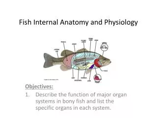
Biology 224 Human Anatomy and Physiology II Week 10; Lecture 1; Monday Stuart Sumida. Human Reproductive Physiology and Reproductive Cycles. OVARY Location : near kidneys, anchored by fallopian tubes to uterus.

E N D
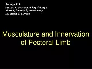
Biology 224 Human Anatomy and Physiology II Week 10; Lecture 1; Monday Stuart Sumida Human Reproductive Physiology and Reproductive Cycles
OVARY Location : near kidneys, anchored by fallopian tubes to uterus. Development: intermediate mesoderm. Ovaries migrate somewhat caudally, retain position near kidneys. Innervation: sympathetic – similar to hindgut, level T12, follows least splanchnic nerve; parasympathetic – sacral outflow Arterial Supply: ovarian artery, branch of abdominal aorta. Venous Drainage: ovarian vein, dump into inferior vena cava.
Ovarian ligament Mesosalpinx Broad ligament
OVARY Function: ovaries produce ova (eggs; singular ovum) in regular cycle determined by hormonal secretions (covered in later lectures). Functions of ovarian hormones and their secretions are tied to secretion of FSH and LH from anterior pituitary gland. ESTROGENS – stimulate development of female sex organs and sexual characteristics. PROGESTERONE + ESTROGENS – regulate menstrual cycle; maintain pregnancy in presence of developing embryo or fetus.
TESTES • Responsible for sperm production and synthesis of male sex hormones. • Location : in postnatal ales, in scrotal sac, connected to inner workings of body by spermatic cord. • Development: from intermediate mesoderm. • As a transitory stage of kidney degenerates, a ligament called the GUBERNACULUM descends on each side of abdomen from inferior pole of gonad. • Gubernaculum passes obliquely through developing anterior abdominal wall at site of future inguinal canal and attaches at internal surface of labioscrotal swelling (future position of scrotum in males or labium majorum in females). • Gubernaculum is thought to guide descent of testes into scrotum, and ultimately anchors testis to scrotal wall.
TESTES Innervation: sympathetic – similar to hindgut, level T12, follows least splanchnic nerve, hook a ride down spermatic cord via testicular blood vessels; parasympathetic – sacral outflow. Arterial Supply: testicular artery. Branches off of abdominal aorta, however developmental proximity ot kidney means they sometimes branch off of renal artery. Arteries follow the developmental track of testes, and can thus be very long. Venous Drainage: testicular vein, dump into inferior vena cava.
TESTES Function: Responsible for sperm production and synthesis of male sex hormones. TESTOSTERONE – stimulate development of male sex organs, secondary sexual characteristics, and behavioral features. Functions of testosterone and its secretion is tied to secretion of LH from anterior pituitary gland.
SPERM AND SPERM PRODUCTION – 1 • Sperm production takes place in the testes. • Each testis contains close to 1000 coiled tubles – SEMENIFEROUS TUBULES – that produce thousands of sperm each second in healthy males. • The inner lining of each tubule is lined with germinal tissue – germinal tissue includes two kinds of cells: • SPERMATOGENIC CELLS – through meiosis, these cells produce haploid sperm cells. All four resultant cells are viable sperm cells. • SUSTENTACULAR CELLS – sustain the spermatogenic cells. They also secrete lubricating fluid to aid outwad movement of sperm as they exit testis via seminiferous tubules and eventually epididymis.
SPERM AND SPERM PRODUCTION – 2 • Between seminiferous tubules are clusters of endocrine cells called LEYDIG CELLS or INTERSTITIAL ENDOCRINOCYTES. • These cells secrete male sex hormones – ANDROGENS, the most important of which is TESTOSTERONE.
SPERM STRUCTURE • Sperm cells are amongst the smallest in the body (~1/20 mm). • Simple construction: head and tail. • Tail is a flagellum – whipping motion provides motility. • Base of tail contains a coiled mitochondrion to provide power for movement. • Head contains nucleus and an organelle called an ACROSOME. • Acrosome contains digestive enzymes that helps sperm to penetrate egg (if present).
EPIDIDYMIS • Seminiferous tubules merge into larger set of tubules called RETE TESTIS. • Rete testis ultimately drains into larger tubules called efferent ducts, which in turn drain into EPIDIDYMIS. • Epididymis includes HEAD, BODY, and TAIL. • Tail of epididymis dilates into DUCTUS DEFERENS.
Sperm are stored at the distal end of the old mesonephric duct...at the distal end of the ductus deferens. This distal end bit that attaches to the testis is called the EPIDIDYMIS.
DUCTUS DEFERENS • DUCTUS DEFERENS passes up spermatic cord, and into body through inguinal canal. • Inside body, right and left ductus deferens pass cranially over ureters, then loops dorsal to them behind the urinary bladder. • As each duct passes behind (dorsal to) bladder, it has appended to it a gland called the SEMINAL VESICLE. • Just prior to attachment of the seminal vesicle, the ductus enlarges into an AMPULLA. • The ampulla is the position of sperm storage prior to ejaculation.
Notice how the spermatic cord loops ventral to (“in front of”) the attachment of the ureter of the bladder.
SEMINAL VESICLE • Plastered up against the dorsal side of urinary bladder. • Exocrine glands -- provide secretions that make up most of seminal fluid. • Fluid lubricates path of exiting sperm. • Fluid is energy-rich (sugar rich) , providing food for sperm. • Secretions slightly alkaline – helps to neutralize slightly acidic environment of vagina. • Once beyond the seminal vesicle, ductus is refered to as te ejaculatory duct.
PROSTATE GLAND • Ejaculatory ducts come together to joint the urethra within the mass of the prostate gland. • Prostate is a single, midline gland just inferior to urinary bladder. • Prostate is a mass of connective tissue, glandular tissue, and smooth muscle. • Prostate secretions: • Fructose • PROSTAGLANDINS – promote uterine contractions to help facilitate sperm movement up uterus into fallopian tubes.
BULBOURETHRAL GLANDS • Pair of glands at base of prostate. • Secretions: • Alkaline pH. • Lubricant for glans of penis.
ERECTION • A PARASYMPATHETIC FUNCTION!! • (A sympathetic reaction doesn’t allow direction of blood to “nonessential” organs.) • Parasympathetic function stimulates dilation of penile blood vessels – causing engorgement of penis with blood. (A hydrostatic skeleton) • Control of Erection (2) • Conscious thoughts (cerebral cortex) stimulate erection center in hypothalamus. This in turn causes vasodilation of penile arterioles. • or • Reflex stimulation of sacral plexus in infants, and sleeping adult males (very common in dream state).
EJACULATION • A SYMPATHETIC FUNCTION!! • Sympathetic fibers innervate smooth muscle of ductus deferens. • Produces forceful peristaltic contractions of smooth muscle of ductus deferens. • Peristalsis propels sperm and seminal fluid out distal end of urethra.
HORMONAL REGULATION IN MALES GONADOTROPIN RELEASING HORMONE (GnRH) – stimulates secretion of Follicle Stimulating Hormone and Luetinizing Hormone. This happens when there is a low concentration of testosterone. FOLLICLE STIMULATING HORMONE (FSH) and LEUTINIZING HORMONE (LH) – both produced by anterior pituitary. Responsible for stimulating spermatogenesis and testosterone secretion. TESTOSERONE – stimulates development of male sex organs, as well as secondary sexual characteristics. Participates in feedback loop involving GnRH. Also inhibits secretion of LH. INHIBIN – secreted by sustentacular cells. Inhibits secretion of FSH.
FEMALE REPRODUCTIVE SYSTEM Human Reproductive Cycles
OVARIES • Remember the following structures: • Mesovarium • Broad ligament • Ovarian ligament • Fimbria • Fallopian tubes
MEIOSIS IN FEMALES • (Recall that in males, each germinal cell produces four haploid cells – each of which becomes a viable sperm cell.) • In females, only one of the resulting cells will be viable and the other three recycled. • After first meiotic division in females, each germinal cell leads to two (diploid). (PRIMARY OOCYTE) • After second meiotic division, the remaining largest cell is the SECONDARY OOCYTE.
FOLLICLES • Each primary oocyte is packaged in an epitheial vesicle called a FOLLICLE. • (It is within follicle that second meiotic division takes place to create secondary oocyte. • Follicular structure has 4 stages: • PRIMORDIAL FOLLICLE (PRIMARY FOLLICLE) – not yet growing. • VESICULAR OVARIAN FOLLICLE (GRAFFIAN FOLLICLE) – about ready to release a secondary oocyte. • CORPUS LUTEUM – what is left of oocyte after it released for ovulation. Corpus luteum secretes ESTROGEN and PROGESTERONE, both of which are important in regulating female menstrual cycle. • CORPUS ALBICANS – degenerate form.
OVULATION • OVULATION is the release of a secondary oocyte from a mature follicle. • Occurs in response to high concentrations of FSH and LH. • Secondary oocyteis “ejected” from ovary directly through mass of ovarian wall. • Fimbria directs oocyte into fallopian tube, preventing movement into coelom.
HORMONAL REGULATION IN NONPREGNANT FEMALE (UTERINE CYCLE)
HYPOTHALAMUS RELEASES GONADOTROPIN-RELEASING HORMONE (GnRH). This stimulates the anterior pituitary to release FSH and LH. • FSH STIMULATES MATURATION OF PRIMARY OOCYTE IN AN IMMATURE FOLLICLE. • FOLLICLE PRODUCES ESTROGEN. Estrogen: (A) builds the uterine wall (the endometrium); (B) inhibits secretion of FSH. • HIGH LEVELS OF ESTROGEN FURTHER STIMULATE SECRETION OF LH BY ANTERIOR PITUITARY. This plus FSH also causes ovulation of the secondary oocyte – leaving follicle without egg (the corpus luteum). • CORPUS LUTEUM SECRETES ESTROGEN AND PROGESTERONE. This maintains the endometrium for 15-16 days and inhibits LH. • (If oocyte is not fertilized and implanted in the uterine wall) CORPUS DEGENERATES (TO CORPUS ALBICANS) AND STOPS PRODUCING ESTROGEN AND PROGESTERONE. • WITHOUT ESTROGEN AND PROGESTERONE, ENDOMETRIUM BREAKS DOWN – MENSTRUATION OCCURS. Menstruation is the sloughing off of the enlarged endometrial wall along with blood and mucous. • DECREASE IN PROGESTERONE AND LH. Low LH causes secretion of FSH by pituitary again. The cycle repeats.
HORMONAL REGULATION IN NONPREGNANT FEMALE • (UTERINE CYCLE) • HYPOTHALAMUS RELEASES GONADOTROPIN-RELEASING HORMONE (GnRH). This stimulates the anterior pituitary to release FSH and LH.
HORMONAL REGULATION IN NONPREGNANT FEMALE • (UTERINE CYCLE) • FSH STIMULATES MATURATION OF PRIMARY OOCYTE IN AN IMMATURE FOLLICLE.
HORMONAL REGULATION IN NONPREGNANT FEMALE • (UTERINE CYCLE) • FOLLICLE PRODUCES ESTROGEN. Estrogen: (A) builds the uterine wall (the endometrium); (B) inhibits secretion of FSH.
HORMONAL REGULATION IN NONPREGNANT FEMALE • HIGH LEVELS OF ESTROGEN FURTHER STIMULATE SECRETION OF LH BY ANTERIOR PITUITARY. This plus FSH also causes ovulation of the secondary oocyte – leaving follicle without egg (the corpus luteum). (Approximately day 15.)
HORMONAL REGULATION IN NONPREGNANT FEMALE • (UTERINE CYCLE) • CORPUS LUTEUM SECRETES ESTROGEN AND PROGESTERONE. This maintains the endometrium for 15-16 days and inhibits LH.
HORMONAL REGULATION IN NONPREGNANT FEMALE • (UTERINE CYCLE) • (If oocyte is not fertilized and implanted in the uterine wall) CORPUS DEGENERATES (TO CORPUS ALBICANS) AND STOPS PRODUCING ESTROGEN AND PROGESTERONE.
HORMONAL REGULATION IN NONPREGNANT FEMALE • (UTERINE CYCLE) • WITHOUT ESTROGEN AND PROGESTERONE, ENDOMETRIUM BREAKS DOWN – MENSTRUATION OCCURS. Menstruation is the sloughing off of the enlarged endometrial wall along with blood and mucous.
HORMONAL REGULATION IN NONPREGNANT FEMALE • (UTERINE CYCLE) • DECREASE IN PROGESTERONE AND LH. Low LH causes secretion of FSH by pituitary again. The cycle repeats.
HORMONAL REGULATION IN NONPREGNANT FEMALE • (UTERINE CYCLE) • IF – SOMEWHERE BETWEEN: • CORPUS LUTEUM SECRETES ESTROGEN AND PROGESTERONE. This maintains the endometrium for 15-16 days and inhibits LH. • And • (If oocyte is not fertilized and implanted in the uterine wall) CORPUS DEGENERATES (TO CORPUS ALBICANS) AND STOPS PRODUCING ESTROGEN AND PROGESTERONE. • SPERM GETS TO EGG... • FERTILIZATION CAN TAKE PLACE, AND ULTIMATELY, EMBRYO CAN BECOME IMPLANTED IN UTERINE WALL.
From point of ovulation (about day 15) to the point where the corpus luteum begins to degenerate (about day 25), fertilization can take place. The potential for fertilization is highest during the first three days of this 10-day period. Sperm with X-chromosome tend to be more robust, and can last longer than those with Y-chromosome. AND, females can be capable of sperm storage. So...if intercourse takes place a bit before ovulation and more robust sperm (with X-chromosome) are stored while weaker (Y-chromosome) sperm die off waiting for ovulation, the chance of having a baby girl increases.
SPERM STORAGE IN THE FEMALE Apparently, females can store sperm up to four days. This explains in part why the “rhythm method” works poorly to avoid pregnancy. (Also, it turns out that unused oocytes are actively scavanged.) With sex before ovulation, sperm can be stored for use. So, even though ovulation hasn’t occurred, pregnancy can occur because the female is holding on the the sperm. Sex AFTER ovulation actually has a slightly lower chance for inducing pregnancy, as the egg could have been scavanged.