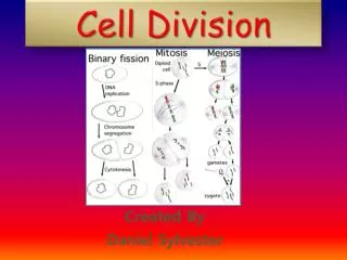
Cell Division
Cell Division. Cell Cycle & Mitosis. Why do cells divide?. DNA Overload – the larger the cell grows, the greater the demands are placed on the cell’s DNA. At some point there is not sufficient DNA for the cell’s proper functioning. Why do cells divide? (continued).

Cell Division
E N D
Presentation Transcript
Cell Division Cell Cycle & Mitosis
Why do cells divide? DNA Overload – the larger the cell grows, the greater the demands are placed on the cell’s DNA. At some point there is not sufficient DNA for the cell’s proper functioning.
Why do cells divide? (continued) Surface Area to Volume Ratio (SA:V) gets too low: As the cell increases in size, the ratio of the surface area to its volume decreases. If this ratio gets too low, the cell cannot efficiently move materials in & out of the cytoplasm.
Mathematical evidence demonstrating S. A. : Vol. Ratio decrease
THE CELL CYCLE Cell cycle is a series of cellular events during which the cell grows, prepares for division & then divides. pg 245- CP text pg 149-H.Bio
INTERPHASE – Made up of the G1, S & G2 phases (Most of the cell life is spent here.) • G1 (Gap 1)- period of cell growth • S (Synthesis) -period of DNA replication • G2 (Gap 2) – period of preparation for cell division. • Cell Division is made up of Mitosis & cytokinesis
Mitosis = division of the nucleus Cytokinesis = division of the cytoplasm Cyclins – proteins that regulate the cell cycle
ALSO: Some cells can stop dividing by exiting the cell cycle and entering a Go phase. There is no preparation for cell division of DNA replication. Central Nervous System cells, like those in the spinal cord are examples of cells that are in the Go phase.
Mitosis Phases & Cytokinesis Mitosis refers to cell division in somatic cells(non sex-cells) PMAT Prophase-Metaphase-Anaphase- Telophase (Interphase: “in-between” period of growth between cell divisions.)
Interphase • DNA –exists in the form of chromatin • Centrioles – a grouping of microtubules that organize the spindle (Not present in plant cells) • During the G2, the chromatin strands change to chromatid pairs (completed in the prophase)
EARLY PROPHASE:(“pro” means before) Chromatin condenses and coils into Chromosomes (consisting of chromatid pairs). This starts at the end of interphase and is completed in prophase. Nuclear membrane breaks down and Nucleolus disappears. Centrioles begin to move to opposite poles of the cell. They eventually reach the ends of the cell. Astral rays appear
Chromosome Structure • When chromosomes form from chromatin, the DNA strands wrap themselves around histone proteins, which help maintain the structure. • The DNA wrapped around histones forms a nucleosome.
Electron micrograph of nucleosomes Each nucleosome contains eight histone proteins (blue), and DNA wraps around these histone structures to achieve a more condensed coiled form.
At the end of G2, (in the beginning of “prophase” in mitosis) the DNA coils up (known as “super coiling”) to become chromosomes. • Coiling of the DNA into chromosomes allows for efficient cell division.
Human Chromosomes • Chromosomes of similar size, shape and containing similar genes are called homologous chromosomes. • Humans have a total of 23 pairs of chromosomes in their body cells.(One of each pair comes from the father, and is called “paternal,” and the other one comes from the mother and is called “maternal”.) • There are 22 pairs of non-sex chromosomes called autosomes, and 1 pair of sex chromosomes.
Diploid v. Haploid number • Diploid = 2N chromosome number = 2 sets of homologous chromosomes. (The diploid chromosome number for human cells is 46.) • Haploid = 1N chromosome number = 1 set of homologous chromosomes. (The Haploid chromosome number for humans sex cells is 23.) Haploid – “Half ”
Late Prophase: Formation of the spindle, composed of fanlike microtubules network that helps to separate the chromosomes. Two types of spindle fibers: polar fibers (pole to pole) kinetochore fibers (attach to centromeres)
Late Prophase: (continued) Chromatid pairs move toward the middle of the cell. (equatorial plane)
Metaphase:(“meta” means middle) • chromatid pairs align themselves in the middle of the cell. • chromatid pairs are held in place by the kinetochore fibers.
Early Anaphase:(“ana” means away) • centromeres split • Spindle fibers are disassembled at the centrioles as the spindle fiber shorten. • chromatid pairs are pulled apart as individual chromatid (now called chromosomes) move to opposite poles. • (Some spindle fibers push against others which help to elongate the cell.)
Late Anaphase • chromosomes are now at opposite poles
Telophase: • chromosomes begin to uncoil into chromatin • cytokinesis (cell splitting) begins as the cytoplasm divides. • The nuclear membrane and nucleolus begin to reappear.
Cytokinesis: Plant and animal cell cytokinesis is different. • Animal Cell -cleavage furrow A constricting actin ring forms around the cell (divides outside –in).
Plant Cell: Cytokinesis • cell plate forms (divides inside-out) It is formed from Golgi vesicles
At the end of cytokinesis there are now two daughter cells in interphase. • They are both diploid (2N) as was the original cell. They are clones of one another.