Download
1 / 19
200 likes | 819 Vues
Learn about the deep fascia of the leg, fascial compartments, anatomy of anterior/lateral leg compartments (muscles, vessels, nerves), and the dorsum of the foot. Explore muscle origins, insertions, actions, and synovial sheaths on the foot.

E N D
ANTERIOR, LATERAL COMPARTMENTS OF THE LEG AND DORSUM OF THE FOOT
OBJECTIVES By the end of the lecture, you should be able to: Identify the deep fascia of leg. Identify the fascial compartments of the leg. Describe the anatomy of the anterior & lateral compartments of the leg (muscles, vessels & nerves). Describe the anatomy and contents of the dorsum of the foot.
The deep fascia surrounds the leg and attached to anterior & medial borders of the tibia. • Two Intermuscular Septa: • Pass from deep aspect of this fascia to be attached to: • Anterior and posterior borders of the fibula(Anterior and posterior fascial septa). • Interosseous membrane: • A thin & strong membrane, that binds the interosseous borders of the tibia & fibula. • It provides attachment for muscles. Fascia of the Leg • membrane:
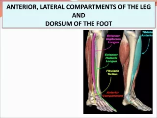
Fascial Compartments of Leg The septa together with the interosseous membrane divide the leg into: Three Compartments: 1. Anterior: Extensors. 2. Lateral: Evertors. 3. Posterior: Flexors. Each compartment has its own Muscles, blood vessels and nerve.
Extensor Retinacula • A thickening band of deep fascia that keeps the long tendons around ankle joint in position. • Superior Extensor retinaculum: • Attached to lower part of anterior borders of tibia & fibula above ankle. • Inferior Extensor retinaculum: • Y-shaped band located anterior to the ankle.
Structures Passing Deep to Extensor Retinacula From medial to lateral: 1. Tom:Tibialis Anterior. 2. Has:Extensor hallucis longus. 3. A:Anterior tibial artery, (ATA) 4. Very: Venae commitant of (ATA). 5. Nice: Anterior tibial nerve, (Deep peroneal nerve). 6. Dog:Extensor digitorum longus. 7. Pig:Peroneus tertius.
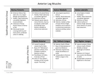
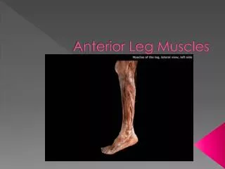
Muscles of the Anterior Compartment • Origin: • All arise from the anterior surface of the shaft of the fibula and interosseous membrane, EXCEPT,tibialis anterior which arises from the lateral surface of the shaft of the tibia and the interosseous membrane.
Insertion & Action of Muscles of Anterior Compartment 1- Tibialis anterior: • Medial cuneiform and • Base of first metatarsal bone. • Action: • Extends (dorsiflexion) of ankle. • Inverts the foot at subtalar joint. • Supports the medial longitudinal arch of the foot. 2- Extensor hallucis longus: • Base of distal phalanx of big toe. • Action: • Extends big toe, • Extends foot at ankle joint; • Inverts foot at subtalar joints.
Insertion & Action of Muscles of Anterior Compartment 3- Extensor digitorum longus: • Extensor expansion of lateral 4 toes. • Action: • Extends foot at ankle joint. • Extends the lateral 4 toes. 4- Peroneus tertius: • Action: • Extends foot at ankle joint. • Everts the foot at subtalar joint.
Synovial Sheaths of Extensor Tendons on the Dorsum of Foot Tibialis anterior and Extensor hallucis longus (Both have their own synovial sheath). Extensor digitorum longus & peroneus tertius: have a common sheath, it extends to the levelof Base of 5th Metatarsal bone.
Lateral Compartment • It contains 2 muscles: • Peroneus longus (PL). • Peroneus brevis (Pb). • Origin: Both arise from the lateral surface of the shaft of the fibula. • Insertion: • PL. Base of first metatarsal & medial cuneiform,(as tibialis anterior). • Pb.Base of fifth metatarsal bone. • Nerve supply: • Both are supplies by superficial peroneal ( Musculocutaneous), nerve.
Lateral Compartment • Action: • Peroneus longus: • Plantar flexes foot at ankle joint; • Everts foot at subtalar joints. • Supports the lateral longitudinal & Transverse arches. • Peroneus brevis: • Plantar flexes foot at ankle joint. • Everts foot at subtalar joint. • Supports the lateral longitudinal arch of foot.
Peroneal Retinacula Superior peroneal retinaculum: Connects the lateral malleolus to calcaneum & holds the tendons of peroneus longus & brevis. Inferior peroneal retinaculum. Synovial Sheaths of Peroneal Longus & Brevis Tendons of the 2 peronei are surrounded by a single common tubular synovial sheath deep to superior peroneal retinaculum. But deep to inferior peroneal retinaculum, each have its separate sheaths.
Deep Fascia of Dorsum of Foot It is very thin, but just distal to ankle joint, it is thickened to form Inferior extensor retinaculum
Extensor Digitorum Brevis • Origin: • Anterior part of upper surface of the calcaneum. • And from inferior extensor retinaculum. • Insertion: • By 4 tendons into the proximal phalanx of big toe. • Extensor expansion of 2nd, 3rd and 4th toes. • Action: • Extend the toes.
Dorsum of Foot • Dorsalis Pedis artery. • Deep & Superficial • Peroneal nerves.
Insertion of Long Extensor Tendons • The tendons of Extensor digitorum longus pass to the lateral four toes. • Each tendon to the 2nd3rd & 4th toes is joined on its lateral side by a tendon of Extensor digitorum brevis. • The extensor tendons form • a Fascial Expansion (Extensor Expansion) on the dorsum of each toe. • The expansion divides into (3) parts. • Central part: inserted into the base of middle phalanx. • Two Lateral parts: inserted into the base of distal phalanx. • The (Extensor Expansion) receives insertion of : • Interossei & Lumbrical muscles.
THANK YOU & BEST WISHES