Download
1 / 51
510 likes | 1.01k Vues
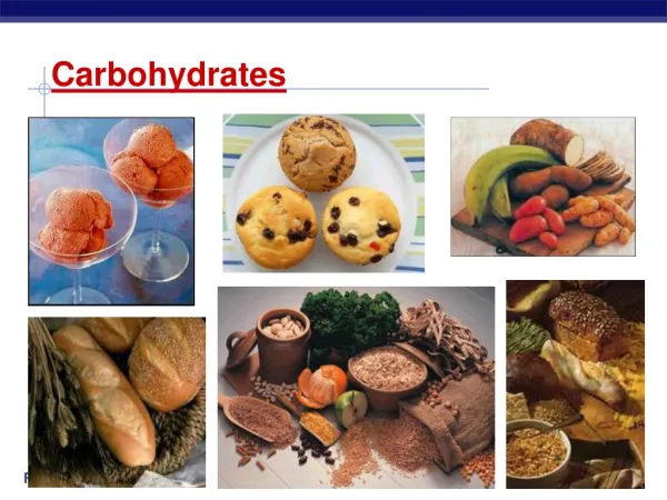
Explore the world of carbohydrates, including their structure, reactions, and analysis techniques. Learn about monosaccharides, glycosides, modified and branched monosaccharides, oligosaccharides, reducing and nonreducing sugars, and carbohydrate composition and linkage analysis. Discover how carbohydrates can be sequenced and the importance of polysaccharides in nature.

E N D
Carbohydrates James R. Ketudat Cairns Aj. Jim Pictures from Stryer, Biochemistry (mostly)
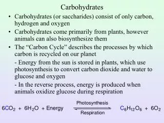
(CH2O)n • Aldehydes (aldose sugars) • Ketones (ketose sugars) • 3 or more carbons.
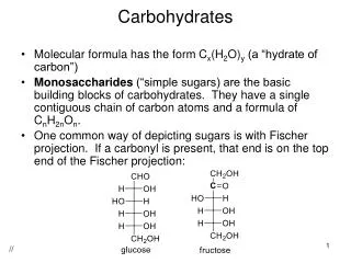
Fischer Projections D and L isostereomers depend on the configuration of the chiral carbon furthest from the carbonyl.
D-Triose to D-Hexose L-sugars are the mirror image of the D-sugar. Sugars that differ in stereochemistry at one position are called epimers.
Carbonyl reactions with alcohols Note: Similar reactions can occur with amines and other nucleophiles.
Monosaccharide cyclization • Formation of an internal hemiacyl or hemiketal is favorable, if it forms a 5 or 6 member ring. • Furanose = 5 member ring • Pyranose = 6 member ring D-Glucopyranose
Anomeric Configuration • Sugars can have two anomeric configurations for each type of ring. • In solution, there are a mix of linear and ring forms that depends on the stability of each.
Sugars are not flat and can form different puckered shapes • Furanose envelopes are most stable. • Pyranose chairs & boats are stable. • Most stable depends on steric interactions. • Axial OH tend to bump, while equatorial do not. Ribose envelopes Glucose chair & boat
Pyranoses can move through many structures, only a few are stable B = boat C = chair H = half chair S = skew boat Vocadlo & Davies, 2008
Glycosides • Reaction at the anomeric carbon (hemiacyl or hemiketal position) form glycosides. • The sugar is trapped in one anomeric configuration. • The bond between the sugar and aglycone is called a glycosidic bond • The product is a glycoside.
Glucosides in nature Glucosides are glycosides with glucose for a sugar. The compounds shown are properly called b-D-glucopyranosides. Ketudat Cairns & Esen, 2010, Cell. Mol. Life Sci.
Modified & branched monosaccharides • Many modified monosaccharides exist in nature. • There are also branched monosaccharides, • e.g. apiose
Oligosaccharides • If two or more monosaccharides polymerize through glycosidic bonds, the are oligosaccharides. • The number of monosaccharides is designated by di-, tri-, tetra-, penta-, hexa- • They can be explicitly described as shown for the common disaccharides to the right. • Often, they are given names like cellobiose, cellotriose or (1,4)-b-D-mannobiose to simply indicate their size and linkage.
Reducing & nonreducing sugars • Sugars that have a free anomeric carbon can undergo redox reactions with Cu2+ • (Fehling’s reagent). • They are called reducing sugars, since they reduce the copper,while they are oxidized.
Redox products of sugars • Sugars can be oxidized at the anomeric carbon to form aldonic acids. • E.g. D-gluconic acid, the produce of a Fehling reagent reaction. • Sugars can be oxidized at a primary alcohol to form a uronic acid. • E.g. D-glucuronic acid, D-galacturonic acid, etc. • These carboxylic acids can form 5 or 6 member rings, such as in L-ascorbic acid. • Sugars can also be reduced to alditols (polyalcohols).
Carbohydrate Composition Analysis • Carbohydrate sugar composition can be tested by hydrolysis (acid or base with heat to break glycosidic bonds), TLC, HPLC, IC or modification and GC/MS. • Compare to standard sugars. • HPLC, IC and GC can potentially quantify sugars. • Modification by acetylation, methylation or trimethyl silanation can make sugars volatile for GC (and acetylation can make them detectable by UV for HPLC).
Carbohydrate Linkage analysis • Carbohydrate linkages can be determined by Nuclear Magnetic Resonance, if the polymer is not too complex. • Methylation analysis can determine which hydroxyls are linked. • First methylate all free hydroxyls • Then hydrolyze glycosidic bonds • Reduce and acetylate the linkage positions. • Run methyl acetyl alditols on GC/MS and compare elution positions to standards. • Does not tell anomeric configuration, just linkage.
Carbohydrate sequencing • Can see the loss of sugars (hexose, pentose, etc.) by mass spectrometry (MS) • Can see fragmentation of sugars in MS spectrum. • Can use specific enzymes to cut off sugars one at a time and look at mass differences. • E.g. neuraminidase to cut off sialic acid, • a-mannosidase to cut off a-linked mannosyl residues. • These enzymes are called glycosidases or glycoside hydrolases (GH).
Positive ion MALDI-TOF mass spectra of derivatized N-linked glycans from bovine fetuin Derivatized with MeI Derivatized with methanol/DMT-MM
Polysaccharides are important structural and storage molecules • Polysaccharides can be grouped by the kinds of monosaccharides they contain • Glucans contain glucose • Mannans contain mannose • Arabinoxyloglucans contain arabinose, xylose and glucose. • Cellulose, a b-glucan is the most abundant polymer on earth. • Chitin/chitosan, a similar structural polysaccharide is also very abundant. • Starch and glycogen represent storage polysaccharides • Alpha-linked glucose polymers
Comparison of Cellulose with Glycogen and Starch • Cellulose is a straight chain, made from alternating orientations of b-1,4-linked glucosyl residues. • Starch and glycogen are coiled a-1,4-linked glucosyl polymers. Amylose coil www.agrana.com/en/1761.asp
Glycogen vs. Starch (Amylopectin) • Glycogen and starch (amylopectin) differ in how many 1,6-linked branches they contain. • Glycogen has an a-1,6-linkage every approx. 8-14 residues. • Starch has a-1,6-linked branches every approx. 24-30 a-1,4-linked residues.
Cellulose in cell wall structure • Cellulose fibers are semicrystalline due to regular hydrogen bonding • Therefore, they are hard to break down.
Plant cell wall polysaccharides • In plant cell walls, the cellulose fibers are linked with hemicellulose (other polysaccharides) and lignin (polyphenolic plastic). Abcbodybuilding.com
Other structural polysaccharides dalwoo.tripod.com/structure.htm
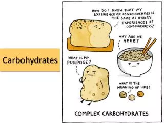
Complex Carbohydrates • Complex carbohydrates are complexes of carbohydrates with other macromolecules • Glycoproteins – found in all domains of life • Proteoglycans • Peptidoglycans (bacterial cell walls) • Glycolipids
Types of Eukaryotic glycoproteins • Cytosolic: single N-acetylglucosamine residues on Ser or Thr hydroxyls. • Likely a regulatory function, like phosphorylation or acetylation. • Secreted: • N-linked: bound to asparagine (Asn, N) • Initial core oligosaccharide added in ER. • O-linked: bound to hydroxyl groups (Ser, Thr, HyPro, HyLys). • Mucin-like added in Golgi • Alpha-mannose linked started in ER • Others
Adding of monosaccharides to molecules • Glycosyl transferases transfer sugars from nucleotidyl glycosides to other molecules in nature. • In the lab, we can also use glycosidases to reverse hydrolyze or transfer glycoside sugars.
Synthesis of core oligosaccharides for N-linked glycosylation • A core oligosaccharide is synthesized on dolichol in the ER membrane for transfer to a glycoprotein Asn in the N-X-S/T sequence. Cytosol ER matrix Dolichol phosphate
Core oligosaccharide addition to proteins • The core oligosaccharide is added to proteins in the ER. • The three Glc residues must be cleaved off before the protein can leave the ER. • Glucose-binding lectins prevent proteins from escaping the ER unfolded. • The alpha-glucosidases that cut off the Glc will not cut off the last until the protein is folded. • If the last Glc is not removed, glc transferase adds another to retain the protein in the ER. • Abnormal O-mannosylation marks for them to be removed from the cycle and degraded.
Calnexin, Glucosidase & Glucosyltransferase ensure secretory protein folding.
C-type lectins like Calnexin use Ca to bind sugars • Lectins are proteins that bind specific sugars • C-type lectins are animal lectins that use bind calcium to help bind the sugar.
Glycosylation is further modified in the Golgi apparatus • In the Golgi glycosidases cut off more of the core oligosaccharides. • Glycosyl transferases add other sugars after the trimming. • The exact carbohydrate varies with the type of organism, cell and protein. • Variation in the amount of carbohydrate added to one protein: microheterogeneity.
Phosphorylation and sulfonation also happen in the Golgi • Phosphomannose is important for sorting of several glycolipid & proteoglycan degrading enzymes to the lysosome. • Lack of the enzymes to transfer the phosphate to mannose results in I-cell disease, where inclusions of undigested glycolipids and proteoglycan develop. • First GlcNAc-Phosphate is added to the Mannose 6-hydroxyl, then GlcNAc is cut off.
O-linked glycoproteins • Secreted O-linked glycoproteins can have from one to thousands of sugars added. • Dystroglycans and some other proteins have alpha-O-Man added in ER. • The first sugar added is usually GalNAc or Gal in mucin-like glycosylation in the Golgi apparatus. • Further sugars added in the Golgi. • The sugars added can contain important information, such as the blood group.
Glycosamino Glycans • Glycosamino glycans are carbohydrates that are usually bound to proteoglycans. • Play important roles in connective tissues. • Also called mucopolysaccharides.
Proteoglycans • Core proteins can have many times their weight in glycosaminoglycan carbohydrate attached. • They hydrate and form a compressible component to give cushioning to joints and related tissues. • They also form a part of the intracellular matrix between cells. Jeffrey & Watt bjr.bjrjournals.org www.histo-moleculaire.com/siteconj/images/037...
Bacterial Peptidoglycans • Peptidoglycans are major components of bacterial cell walls • Thick coating on outside of Gram positive bacteria • Thinner layer between membranes of Gram negative bacteria • Cut by Lysozyme. Gram positive bacteria cell wall structure
Peptidoglycan cell walls Staphylococcus aureus peptidoglycan
Glycolipids Glycolipids: Glycosphingolipids Glycoglycerol lipids: Plant galactolipids Animal PtdGlc (below) Cholesterol glucoside Wennekes et al., 2009, Angew. Chem. Int. Ed. 48, 8848-8869 Ishibashi et al., 2013
Glycosphingolipid synthesis & catabolism Wennekes et al., 2009, Angew. Chem. Int. Ed. 48, 8848-8869
Carbohydrate Active proteins • Carbohydrate binding proteins/domains • Carbohydrate binding modules (CBM) can bind simple sugars or more extensive regions. • Lectins bind simple sugars. • Carbohydrate Active Enzymes (CAZy) • Glycoside Hydrolases (GH, glycosidases) • Transglycosidases (TG) catalyze transfer rather than hydrolysis. • Glycosyl Transferases (GT) • Polysaccharide Lyases- nonhydrolytic cleavage of glycosyl linkages • Carbohydrate Esterases • Other carbohydrate modifying enzymes
Viral Carbohydrate-Active Proteins • The flu virus strains are distinguished by forms of carbohydrate active proteins. • Hemaglutanin (H in virus name) binds to sialic acid on cell surface to invade. • Neuraminidase (N in virus name) cuts off the sialic acid to free the virus, once inside the cell.