Download
1 / 30

E N D
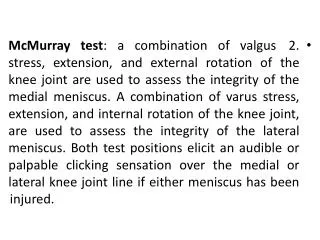
2. McMurray test: a combination of valgus stress, extension, and external rotation of the knee joint are used to assess the integrity of the medial meniscus. A combination of varus stress, extension, and internal rotation of the knee joint, are used to assess the integrity of the lateral meniscus. Both test positions elicit an audible or palpable clicking sensation over the medial or lateral knee joint line if either meniscus has been injured.
Fig. 2 . The McMurray test for meniscal tears. flexed, internally and externally the knee. rotate the tibia on the femur.
the test is considered positive for a torn medial meniscus, usually in the posterior position. With the leg externally rotated and in valgus, slowly extend the knee. If click is palpable or audible, With the leg externally rotated, place a valgus stress on the knee.
3- The medial-lateral grind test: from a starting position of full extension, alternating varus and valgus stresses are applied as the knee is alternately flexed and extended to and from a position of 45 degrees flexion. A longitudinal meniscal tear produces a grinding sensation along the lateral or medial joint line according to the involved side. Prolonged grinding indicates a complex meniscal tear or injury of both menisci.
Bragard sign This test may be used if anterior joint-line point tenderness is present . To test for a medial lesion, the examiner extends and externally rotates the tibia, which displaces a meniscal lesion forward, if one exists. Palpable tenderness along the anterior medial joint line is reduced with flexion and internal rotation
Bounce home test The patient is supine with his heel cupped in the examiner's hand . The examiner fully flexes the knee and then passively extends the knee. If the knee does not reach complete extension or has a rubbery or springy end feel, the knee movement may be blocked by a torn meniscus .
Merkel sign • Instruct the patient to stand with his knees extended and to rotate the trunk. This movement causes compression of the menisci. • Medial compartment pain during internal rotation of the tibia indicates a medial meniscal lesion. Lateral compartment pain occurring during external rotation of the tibia indicates a lateral meniscal lesion.
Payr sign • With the patient sitting cross-legged, the examiner exerts downward pressure along the medial aspect of the knee. • Medial knee pain indicates a posterior horn lesion of the medial meniscus
First Steinmann sign • With the patient supine and the knee and hip flexed at 90°, the examiner forcefully and quickly rotates the tibia internally and externally. • Pain in the lateral compartment with forced internal rotation indicates a lateral meniscus lesion. Medial compartment pain during forced external rotation indicates a lesion of the medial meniscus.
Second Steinmann sign • This test is indicated when point tenderness is located along the anterior joint line. • When the examiner moves the knee from extension into flexion, the meniscus is displaced posteriorly, along with its lesions. The point of tenderness also shifts posteriorly toward the collateral ligament.
Operative management • The overall treatment goal is to preserve as much meniscal tissue as possible while addressing the clinical symptoms caused by the meniscal tear. • Meniscal tears in the outer third or vascular zone will heal and therefore a meniscal repair is recommended. • Meniscal tears that extend beyond the outer third or vascular zone will not heal and therefore a partial meniscectomy is recommended. • A complete meniscectomy may be performed especially with significant degenerative tears to the meniscus. • A partial meniscectomy leaves a rim of tissue in place, which maintains some stress protection for the articular cartilage, in contrast to a total meniscectomy, which (in the absence of regeneration) is associated with increased cartilage degeneration, joint narrowing, alterations in bone geometry, and osteophyte formation.
Surgical procedure: the procedure is almost universally done today by arthroscopic means. Partial meniscectomy is indicated in unstable tears that are not repairable due to location or configuration and In this procedure, the surgeon removes only the damaged or unstable portion of the meniscus, and balances the residual meniscal rim. • Specifically, removal of the menisci nearly doubles the articular cartilage stress on the femur and multiplies the forces by six or seven times on the tibial plateau. The increase in joint stress may contribute to degenerative changes within the tibiofemoral joint. • The procedure for a total meniscectomy, the entire meniscus may be removed.
Preoperative rehabilitation • Pre operative rehab for a meniscal injury that is to undergo a meniscectomy may involve: • (1) Swelling and pain control • (2) Range of motion exercises • (3) Quadriceps strengthening • (4) Aquatic therapy for strengthening if pain is preventing strengthening with normal weight bearing
POSTOPERATIVE REHABILITATION • Goals 1- Control of pain and edema 2- Obtaining and maintaining full ROM 3- Regaining proper quadriceps strength. 4- Immediate weight bearing as tolerated 5- Return to activity
Phase 1: Acute phase (Maximum protection phase first 10 days) • Goals: 1- Diminish Swelling and pain 2- Improve ROM 3- Reestablish quadriceps muscle activity
Phase 1: Acute phase (Maximum protection phase) • Intervention: Days 1-3 • The patient is subjected to CPM and also fitted with a motion-control brace. Knee motion is restricted for both modalities as follow according to the site of lesion: 0 to 90 degrees knee flexion in case of peripheral defect in the middle portion of the meniscus, 20 to 90 degrees with a peripheral defect in either the anterior or posterior horn of the meniscus, and 20 to 70 degrees flexion if the defect is in the central nonvascular region. • • Cryotherapy • • Light compression wrap • • Electrical muscle stimulation to quadriceps • • Active assisted ROM stretching, emphasizing full knee extension (flexion to tolerance) • • Strengthening Exercises: Straight leg raises, hip adduction and abduction, ¼ and/or ½ squats Weight bearing as tolerated (partial use of axillary crutches)
Days 4-7 • • Cryotherapy and continued use of compression wrap • • Electric muscle stimulation to quadriceps • • Active assisted, passive ROM, and stretching exercises (hamstrings, calf muscles, quadriceps) • • Strengthening Exercises: Straight leg raises, quadriceps sets, hip adduction and abduction, knee extension 90-40 degrees, ¼ and/or ½ squats • • Weight bearing (partial weight bearing)
Days 7-10 • • Continue all exercises and add: Leg press (light weight), toe raises, and hamstring curls • • Bicycle (when ROM 0-105 degrees with no swelling)
Phase 2: Internal Phase (Moderate protection phase) • Goals: 1- Reestablish full non painful ROM 2- Restore and improve muscular strength and endurance 3- Proprioceptive training 4- Gradual return to functional activities
Phase 2: Internal Phase (Moderate protection phase) • Intervention: • Days 10-17 • • Stretching exercises • • Strengthening exercises: Lateral lunges, front lunges, ½ squats, leg press, lateral step ups, knee extension (90-40 degrees), hamstring curls, hip adduction and abduction, hip flexion and extension, toe raises • • Proprioceptive and balance training • • Bicycle, Stairmaster • Full weight bearing as tolerated
Day 17-Week 7 • • Continue all exercises • • Pool program (deep water running and leg exercises) • Maximal isotonic full range activities. • Isokinetic exercises. • Flexibility procedures to ensure normal ROMS of knee and patellofemoral joints. • Proprioception training is continued. • Closed kinetic chain exercises (neuromuscular integration). • Cardio-vascular and cardio-muscular endurance is emphasized by and including swimming. • Extended walking.
Criteria for progression to Phase 3: Satisfactory clinical examination (minimal effusion) • Full/non painful ROM • No pain or tenderness • Satisfactory isokinetic test
Phase 3: Advanced Activity Phase (Return to activity phase) • Weeks 7-12 • Goals: 1- Maintain full ROM 2- Enhance muscular strength and endurance 3-Return to sport/functional activities
Intervention: • • Therapeutic exercises: Continue to • May begin plyometrics • Begin running program and agility drills • Gradually return to sports competition
Criteria for Return • The athlete may return to activity when (1) Swelling does not occur with activity. (2) Full ROM has been regained, (3) There is equal bilateral strength in knee flexion and extension, (4) The athlete can successfully complete functional performance tests.