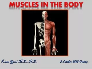
Kaan Yücel M.D., Ph.D
220 likes | 439 Vues
Posterior aspect of the forearm & Anatomy of the hand. IN 19 QUESTIONS. Kaan Yücel M.D., Ph.D. 12.January.2012 Thursday. THE ANATOMY OF. POSTERIOR ASPECT OF THE FOREARM. IN. 7 QUESTIONS. 1. How are the muscles in the posterior compartment estabslihed?. In two layers:

Kaan Yücel M.D., Ph.D
E N D
Presentation Transcript
Posterior aspect of the forearm • & • Anatomy of the hand • IN 19 QUESTIONS • Kaan Yücel M.D., Ph.D • 12.January.2012 Thursday
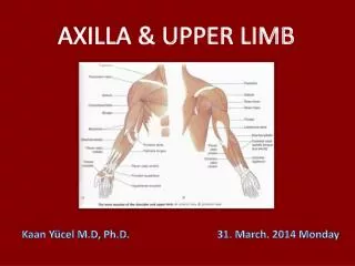
THE ANATOMY OF POSTERIOR ASPECT OF THE FOREARM IN 7 QUESTIONS
1. How are the muscles in the posterior compartment estabslihed? In two layers: a superficial layer a deep layer.
2. Which muscles are in the superficial layer? Lateral epicondyle of humerus Brachioradialis Extensor carpi radialis longus Supraepicondylar ridge of humerus Supraepicondylar ridge of humerus Lateral epicondyle of humerus (common extensor origin) Extensor carpi radialis brevis Lateral epicondyle of humerus (common extensor origin) Lateral epicondyle of humerus; posterior border of ulna flexion of forearm extendand abduct hand at the wrist joint Extensor carpi ulnaris Extends and adducts hand at wrist joint assists triceps in extending forearm stabilizes elbow joint; may abduct ulna during pronation Lateral surface of olecranon and superior part of posterior surface of ulna extends medial four digits primarily at metacarpophalangeal joints, secondarily at interphalangeal joints Extensor digitorum Extend and abduct hand at the wrist join Extensor digiti minimi Extensor expansion of 5th digit Anconeus Extensor expansions of medial four digits Lateral surface of distal end of radius proximal to styloid process Base of 3rd metacarpal Base of 5th metacarpal Base of 2nd metacarpal Base of 3rd metacarpal Base of 5th metacarpal Base of 2nd metacarpal Common origin from the supraepicondylar ridge and lateral epicondyle of the humerus Except for the brachioradialis and anconeus, extend as tendons into the hand.
3. Which muscles are in the deep layer? Supinator Abductor pollicis longus Extensor pollicis brevis Extensor pollicis longus Extensor indicis Except for the supinator muscle, all these deep layer muscles originate from the posterior surfaces of the radius, ulna, and interosseous membrane and pass into the thumb and fingers.
4. What movements do these muscles do? EXTENSION OF THE HAND & FINGERS ABDUCTION OF THE FINGERS SUPINATION
5. How are the muscles of the posterior compartment of the forearm innervated? Supinator Extensor carpi radialis brevis Deep branch of radial nerve Rest Posterior interosseous nerve continuation of deep branch of radial nerve
6. ....arteries in the posterior compartment of the forearm? Radial artery Posterior interosseous artery origin: common interosseous branch of the ulnar artery recurrent interosseous artery End by joining to dorsal carpal arch of the wrist Anterior interosseous artery origin: common interosseous branch of the ulnar artery
7. ...radial nerve in the posterior compartment of forearm? Deep branch becomes posterior interosseous nerve after emerging from between 2 heads of supinator Posterior interosseous nerve passes deep to extensor pollicis longus to reach the wrist.
THE ANATOMY OF THE HAND IN 12 QUESTIONS
2. What are flexor retinaculum & carpal tunnel? The carpal tunnelformed anteriorly at wrist by a deep arch formed by carpal bones & flexor retinaculum (transverse carpal ligament) Flexor digitorum/superficialis Flexor pollicis longus Median nerve Pass through the carpal tunnel
3. What is extensor retinaculum (dorsal carpal ligament)? The extensor tendons pass into the hand in six compartments defined by an extensor retinaculum: • extensor digitorum & extensor indicis posterior surface of the wrist extensor carpi ulnaris & extensor digiti minimi medial side of the wrist • abductor pollicis longus & extensor pollicis brevis • extensor carpi radialis longus & extensor carpi radialis brevis • extensor pollicis longus • through three compartments on the lateral surface of the wrist.
4. What is palmar aponeurosis? A triangular condensation of deep fascia that covers the palm and is anchored to the skin in distal regions. Continuous with the palmaris longus tendon, when present; otherwise, anchored to the flexor retinaculum.
5. What are extensor hoods for? Tendons of the extensor digitorumextensor pollicis longus musclesexpand over the proximal phalanges to form "extensor hoods" or "dorsal digital expansions". Tendons of the extensor digiti minimi, extensor indicis, extensor pollicis brevisjoin these hoods.
6. Which are the intrinsic muscles of the hand? Palmaris brevis Thenar muscles Abductor pollicis brevis Abductor digiti minimi Opponens pollicis Hypothenar muscles Flexor digiti minimi Adductor pollicis Flexor pollicis brevis Lumbrical Interossei
7. How are the intrinsic muscles innervated? All of the intrinsic muscles of the hand by deep branch of the ulnar nerve Except three thenar & two lateral lumbrical muscles by median nerve
8. ....arteries of the hand? Superficial palmar arch Deep palmar arch
9. ....veins of the hand? Cephalic veinoriginates from lateral side of dorsal venous network. Basilic vein originates from medial side of dorsal venous network.
10. ...sensory innervation of the hand? Ulnar nerve medial side of the palm,medial half of the dorsum of the hand, the 5th finger, and the medial half of the 4th finger,anterior surfaces of the medial one and a half digits, Median nerve thumb,index,middle fingers,lateral side of the ring [distal parts on the dorsum of the hand] Radial nerve dorsolateral side
11. ...carpal tunnel? Homework: • 1. Which structures pass through the carpal tunnel and their anatomical relationships with each other in the tunnel? • 2. The incidence of carpal tunnel syndrome in the world and/or in Turkey? • 3. The risk factors, higher in whom? Any gender disperancies in its incidence? Please send answers to yeditepeanatomy@yahoo.com