Download
1 / 30
300 likes | 455 Vues
CELLULAR SIGNAL TRANSDUCTION 2) From the membrane to the nucleus TEIL F TRANSCRIPTION FACTORS AND THE REGULATION OF TRANSCRIPTION Rainer deMartin / Erhard Hofer Institute of Vascular Biology and Thrombosis Research Vienna Competence Center Lazarettgasse 19, 1090 Wien. Erhard Hofer

E N D
CELLULAR SIGNAL TRANSDUCTION 2) From the membrane to the nucleus TEIL F TRANSCRIPTION FACTORS AND THE REGULATION OF TRANSCRIPTION Rainer deMartin / Erhard Hofer Institute of Vascular Biology and Thrombosis Research Vienna Competence Center Lazarettgasse 19, 1090 Wien
Erhard Hofer activation of transcription factors by surface receptors Summary selected examples CREB, SRF, NFAT, SMAD additional specific example: - Regulation of signaling pathways/transcription factors by proteolytic cleavage: WNT, NOTCH (embryonic development, adult stem cells) Nuclear import, -export Chromatin Transcription initiation complexes Rainer deMartin: Principals of transcriptional regulation Structural features of transcription factors Basic mechanisms of transcriptional regulation
Signaling pathways: Receptor to transcription factors SMAD Ras A-Cyclase PLC STAT STAT IKKK SMAD/Co-SMAD cAMP PKA NFAT / NFkB STAT SRF SMAD
Gene regulation by PKA: CREB bound to CRE Is phosphorylated by PKA, activates transcription without effect on DNA binding Example 3
The phosphorylated MAPK ERK is transported into the nucleus and phosphorylates the transcription factor TCF on the promoter or: PLC-g Raf MEK Genes for Cell cycle/ Proliferation early response genes, c-fos ERK: extracellular signal regulated kinase TCF: ternary complex factor SRF: serum response factor SRE: serum response element (DNA binding sequence for TCF and SRF in promoter of various genes)
Ca++ Signaling pathway - Gene regulation the Phosphatase Calcineurin dephosphorylates NFAT NFAT translocates Into nucleus NFAT= transcription factor (nuclear factor activated T cell) Ca++ P I NFAT Calmodulin Calcineurin P Kern
Transport of phosphorylated SMADs into nucleus
Regulation of transcription factors by proteolytic cleavage
WNT signaling pathway Secreted signaling peptide, important in embryonic development Mutatios on proteins of wnt signaling pathway frequent in cancer Wnt Wingless (Drosophila) Int-1 (breast cancer oncogen) (detected experimentally by virus Integration) In signalling pathway: APC (adenomoteous polyposis coli) mutated in adenoma of colon and 80 % of colon cancer induces myc gene and proliferation Example 4a
b-Catenin signaling pathway: w/o signal: b-catenin is continously phosphorylated, ubiquitinylated, degraded in proteasom Wnt-signal: Kinase is inhibited, non-phosphorylated b-Catenin transported into nucleus, aktivates transcription by competing of a corepressor (LDL rceptor related protein) (Signaling protein) Phosphorylation, Ubiquitinylation, degradation in proteasom (b-Catenin Coaktivator) (Corepressor)
components of the WNT signaling pathways e.g. important for maintaining the stem cell population in gastro-intestinal tract, Over-activation by APC mutation - cancer
Notch hedgehog example 5b 1- Embryonic development E.g. nerve cells Drosophila, Delta on nerve cells signals to neighbouring cell: Do not differentiate to nerve cell, Peptide translocates into nucleus and converts CSL to become an activator 2- Angiogenesis: Tip versus stalk cell, tip cell signals stalk cell not to become another tip cell
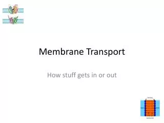
Nuclear membrane: Double membrane with nuclear pores
Nuclear pore complex innere Seite
The nuclear localization signal is a basic amino acid sequence Model of nuclear import Mediated by the small G-protein Ran
Summary Interphase Metaphase
HAT HDAC condensed CHROMATIN no TRANScRIPTION Z.B. HETEROCHROMATIN loose CHROMATIN TRANCRIPTION Z.B. EUCHROMATIN Ac Ac CHROMATIN NUCLEOSOM DNA HAT = HISTONACETYLTRANSFERASE ACTIVATOR OF TRANSCRIPTION HDAC = HISTONDEACETYLASE REPRESSOR of TRANSCRIPTION GENEXCS18
HDAC HAT REGULATION OF TRANSCRIPTION BY CHANGE OF CHROMATIN STRUCTUR REPRESSION Repressor X Target GENE condensedChromatin Aktivator ACTIVIERUNG Z Y A Target GENE openChromatin GENEXCS19
Mediator DNA Looping