Download

1 / 20
210 likes | 622 Vues
Organic Chemistry , 6 th Edition L. G. Wade, Jr. Chapter 13 Nuclear Magnetic Resonance Spectroscopy. Jo Blackburn Richland College, Dallas, TX Dallas County Community College District ã 2006, Prentice Hall. Introduction.

E N D
Organic Chemistry, 6th EditionL. G. Wade, Jr. Chapter 13Nuclear Magnetic Resonance Spectroscopy Jo Blackburn Richland College, Dallas, TX Dallas County Community College District ã 2006,Prentice Hall
Introduction • NMR is the most powerful tool available for organic structure determination. • It is used to study a wide variety of nuclei: • 1H • 13C • 15N • 19F • 31P => Chapter 13
The NMR Spectrometer => Chapter 13
Old School NMR Chapter 13
High Tech NMR Chapter 13
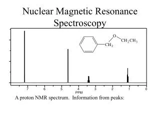
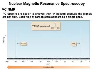
NMR Signals • The number of signals shows how many different kinds of protons are present. • The location (chemical shift) of the signals shows how shielded or deshielded the proton is. • The intensity of the signal shows the number of protons of that type. • Signal splitting shows the number of protons on adjacent atoms. => Chapter 13
How Many Kinds of Protons • Depends on symmetry and chemical environment • The number of signals is equivalent to the number of different kinds of protons Chapter 13
=> Protons in a Molecule Depending on their chemical environment, protons in a molecule are shielded by different amounts. Chapter 13
The NMR Graph 2 1 3 => Chapter 13
Chemical Shift • Measured in parts per million. • Ratio of shift downfield from TMS (Hz) to total spectrometer frequency (Hz). • Same value for 60, 100, or 300 MHz machine. • Called the delta scale. => Chapter 13
Location of Signals • More electronegative atoms deshield more and give larger shift values. • Effect decreases with distance. • Additional electronegative atoms cause increase in chemical shift. => Chapter 13
Typical Values => Chapter 13
O-H and N-H Signals • Chemical shift depends on concentration. • Hydrogen bonding in concentrated solutions deshield the protons, so signal is around 3.5 for N-H and 4.5 for O-H. • Proton exchanges between the molecules broaden the peak. => Chapter 13
The NMR Graph 2 1 3 => Chapter 13
Spin-Spin Splitting • Nonequivalent protons on adjacent carbons have magnetic fields that may align with or oppose the external field. • This magnetic coupling causes the proton to absorb slightly downfield when the external field is reinforced and slightly upfield when the external field is opposed. • All possibilities exist, so signal is split. => Chapter 13
1,1,2-Tribromoethane Nonequivalent protons on adjacent carbons. => Chapter 13
The N + 1 Rule If a signal is split by N equivalent protons, it is split into N + 1 peaks. => Chapter 13
Range of Magnetic Coupling • Equivalent protons do not split each other. • Protons bonded to the same carbon will split each other only if they are not equivalent. • Protons on adjacent carbons normally will couple. • Protons separated by four or more bonds will not couple. => Chapter 13
Splitting for Ethyl Groups => Chapter 13
Splitting for Isopropyl Groups => Chapter 13