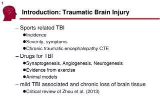
Introduction: Traumatic Brain Injury
440 likes | 640 Vues
Introduction: Traumatic Brain Injury. Sports related TBI Incidence Severity, symptoms Chronic traumatic encephalopathy CTE Drugs for TBI Synaptogenesis, Angiogenesis, Neurogenesis Evidence from exercise Animal models mild TBI associated and chronic loss of brain tissue

Introduction: Traumatic Brain Injury
E N D
Presentation Transcript
Introduction: Traumatic Brain Injury • Sports related TBI • Incidence • Severity, symptoms • Chronic traumatic encephalopathy CTE • Drugs for TBI • Synaptogenesis, Angiogenesis, Neurogenesis • Evidence from exercise • Animal models • mild TBI associated and chronic loss of brain tissue • Critical review of Zhou et al. (2013)
TBI in the USA • 1.7 Million/year • 29% of all ER visits • Incidence ‘bathtub’ function. 2002-2006 Faul et al. (2010)
TBI in the USA • Causes • Also vary with age
Sports related TBI • CDC estimates 300,000 sports related TBIs each year. • However, only included TBIs with loss of consciousness. • LoC only account for 8-19.2% of of sports related TBIs. • Therefore, ~1.6-3.8 million sports related TBIs each year. • Athletes tend to under-report: Even this estimate may be low! Langlois et al. (2006)
Sports concussion • Recent attempts to define sports-related sub-classification of mild TBI (e.g. Cantu Grading, American Academy of Neurology) • Often no loss of consciousness. • Post-traumatic amnesia and mental status can appear normal within minutes.
Sports concussion • Most athletes report complete resolution within 5-10 days. • ‘Post-Concussive Syndrome’ refers to complaints that persist weeks to months – more common with multiple events. • According to ICD-10 and DSM-IV mild TBI can occur without loss of consciousness, but PCS requires LoC. • While some individuals who claim chronic problems may be malingering, others with legitimate problems may not get appropriate compensation.
Return to play • Strong reasons to halt play after mTBI: • Concussion leaves one susceptible to another, especially if second impact occurs before symptoms resolve. • Progressive process: smaller impacts cause the same symptom severity (Zurich statement). • Repeated concussions may increase the risk in later life for dementia, Parkinson's disease, and/or depression.
Return to play • McCrea et al. (2003) examined 1631 collegiate football players – carefully evaluated 94 who suffered concussion (and 56 controls) 3hr, 1,2,3,5,7,90 days post injury. Cognitive deficits cleared in 5-7 days, balance deficits 3-5 days. Verbal processing resolved over 7 days.
Return to play • Quigley’s rule (Schneider, 1973) termination of contact sports after 3 concussions, regardless of severity • Zurich Statement (2008) provides graduated return of activities, typically over one week (table 1), with recognition of modifiers (table 2). • Section 4.2 same day return to play when concussion management team available (professional sports)
Second Impact Syndrome • SIS refers to fatal injury that results occurs if two mild TBIs occur in short succession (Buzzini & Guskwicz, 2006; Solomos 2002). • Possible mechanism: first injury impairs vascular regulation (Hovda et al., 1999). • SIS also referred to as diffuse cerebral swelling (DCS) • SIS very rare (17 documented cases), so remains controversial (McCory 2001), unlikely well documented vulnerability to subsequent non-fatal mild TBI.
Chronic traumatic encephalopathy • “Punch drunk syndrome” (Martland 1928) • “Dementia Pugilistica” (Lampert and Hardman, 1984) • CTE (Modern) • Professional US Football conservatively 3.7% incidence (Gavett et al., 2011). Some evidence for wrestling, hockey. • Main symptoms typically years after career. • Histological signs can be seen early in life, e.g. University player Owen Thomas who committed suicide at 21 years old. • Seen in animals and humans with single blast exposure.
CTE, McKee et al 2013 • Recent histological study of 85 men (17-98yo) with history of repetitive mild TBI and 18 controls: 68/85 had evidence of CTE. • 63% CTE only • 16% Lewy Body disease • 12% motor neuron disease (Lou Gherig’s, ALS) • 11% Alzheimers • 6% Frontotemporal degeneration • Authors suggests progression of tau abnormalities Lou Gherig
Histological Biomarkers for CTE • Tau-positive neurofibrillary tangles (NFTs) in the neocortex, concentrated around penetrating parenchymal vessels • Neuropil (cortical) threads • Neocortical diffuse amyloid plaques, with or without neuritic plaques • Sparing of the hippocampus
AD vs CTE (McKee et al., 2013) AD • Neurofibrillary tangles (expressing amyloid, red), normal sulcal depth. • No clustering of tangles near blood vessels • Diffuse tau CTE • Clumps of tau, large sulci • Clustering of tangles near blood vessels • Tau mostly in superficial layers
CTE : McKee et al., 2013 • Currently CTE diagnosed at autopsy. Symptoms very similar to Alzheimer’s disease • Depression, anger around 35 yo • Of 36 former athletes, 6 committed suicide • Mental decline around 59 yo • General symptoms • Recurrent headaches • Dizziness • Mood disorders (depression) • Aggression • Impaired judgment and impulse control • Parkinsonian movement disorders • Progressive dementia • Very strong association with ALS
Genetic Risk Factors • ApoE E4 a strong risk factor for Alzheimer’s disease. • E2: reduced AD risk • E3: normal AD risk • E4 single copy: x1.75 risk • E4 two copies: x8 risk • May also be involved with sports-related dementia: • Jordan et al. (1997) boxing exposure and E4 interacted to predict dementia. • Kutner et al. (2000) cognitive effects in US Football players with E4.
Summary of Sports-related TBI • Sports related mTBI very common. Boxing, football, ice hockey, soccer (also equestrian, rugby and gymnastics). • Reported incidence increasing (better awareness). • Both acceleration/deceleration impacts as well as rotational shearing of white matter. • Typically not visible with neuroimaging • Basal forebrain, medial temporal lobes, white matter. • Typically 5-10 day recovery
Break -Pause
Potential for treating TBI • How can we reduce the consequences of TBI? • Neurogenesis (Chapter 20) • Xiong et al. (2010) review restorative treatments. • Xiong et al. (2013) describe animal models of TBI.
Can drugs treat TBI? (ch 20) • Primary Injury • Direct due to mechanical forces - contusion, damaged blood vessels, axonal shearing. • Secondary Injury • Cascade of metabolic cellular(e.g. increased Ca,Na decreased K) and molecular events leading to tissue damage. • e.g. Glutamate toxicity, perturbed calcium homeostatis, increased free radicals lipid peroxidation, mitochondrial dysfunction (due to Ca), inflammation, apoptosis (programmed cell suicide) and diffuse axonal injury. • Initial contusion hours/days: swollen, days-weeks: shrunken (pyknosis: nucleus shrinks) and resorbed GM and WM.
Acute and chronic injury • Primary and secondary Xiong, Mahmood & Chopp (2010)
Protecting people from TBI • Both Primary and Secondary Injury • Prevention: seatbelts, helmets • Secondary injury • Time window to intervene with processes • Some studies suggest long term functional and structural changes take place up to 1 year post injury. • Drugs taken after injury may help minimize extent of injury (neuroprotection) and aid in compensation (neurorestoration).
Neuroprotection versus neurorestoration • Neuroprotective therapies aim to block the molecular cascade of injury following traumatic brain injury (TBI). • These approaches aim to minimize size of injury. • To date, human clinical data disappointing. • Alternatively, neurorestoration therapy aims to aid recovery via neurogenesis, axonal sprouting, synaptogenesis, oligodendrogenesis and angiogenesis Xiong, Mahmood & Chopp(2013)
Mechanisms for restoration • Adult CNS has limited capacity to regenerate after injury (Xiong et al., 2010). • Neurogenesis • Angiogenesis • Axonal sprouting • Glial responses • Synaptic plasticity appears dominant mode of adaptation in adult brain (includes biochemical changes and synaptogenesis)
Neurogenesis • Adults develop new neurons in and around the hippocampus. • TBI induces neurogenesis in mice (Kernie et al., 2001) • Wang et al (2001) report Metformin promotes mouseneurogenesis and enhancesspatial learning
Neurogenesis (ch 20) • In chapter 20, Wojtowicz suggests • Hippocampal neurogenesis first described in 1962 (Altman) • ‘Use it or lose it’ seems to apply • Some initial examples of adult neurogenesis in other brain regions. • The authors suggest that neurogenesis may be different for lab animals versus those in natural environment. • Weak evidence: they compare across species (rats vs squirrels) • Studies within species (rats, Epp et al., 2009) does not support this claim • Unlike embryonic neurogenesis, adult neurogenesis appears to be limited (Kriegstein and Alvarez-Buylla, 2009): • Only adds to existing cortical layers • Only limited brain areas (hippocampal, olfactory bulb) • New cells migrate through existing layers • Derived from specialized glial rather than stem cells • Only generates specific types of neurons
Angiogenesis • Vasculature can grow to provide more nutrients. • Most prior research has focused on drugs that impair angiogenesis (to treat tumors) • Meng et al. (2011) note EPO given 24 hours after TBI increase angiogenesis and hippocampal neurogenesis in rats.
Axonal Sprouting (Axonal Remodeling) • Regeneration of axons well known in peripheral nervous system. • Recently: CNS axonal sprouting may be involved with motor recovery after TBI (Smith et al., 2007; Oshima et al., 2009) • Inosine infused into ventriclesmay aid this (Smith et al., 2007)
Glial responses to TBI • Gliosis common after injury • Whereas adult neurogenesis is focal/limited, glial responses widespread/robust. • Astrocytes react to TBI, with response graded by injury severity. Both beneficial and detrimental consequences • Detrimental: numerous studies (e.g. Rodgers et al., 2013) find that anti-inflammatory (that reduce glial response) help recovery. • Beneficial: Myer (2006) in mice with moderate TBI, selective ablation of the reactive astrocytes increased neuronal degeneration by 60% Astrocytes react to injury Ren et al. (2013)
Synaptic plasticity • Existing synapses’ facilitory and inhibitory connections strengthen and weaken over time. • Dynamic and rapid method for learning and compensation (Hebbian learning). • Albensi et al. (2000) gave rats cyclosporin A (a compound that stabilizes mitochondrial function) 24 hours after TBI and found normalized long-term potentiation and normal long-term depression.
Drug intervention • Multiple mechanisms of injury • Cocktail of drugs may be required. • Unfortunately, drug interactions make these studies challenging • Example: EPO promising found promising for minimizing ischemic stroke, but clinical studies found interaction with tPA (clot busting agent). Xiong, Mahmood & Chopp (2013)
Animal models of TBI • Human injury very heterogeneous – hard to conduct controlled studies. • Different forms of animal injury attempt to mimic specific patterns of human injury. Xiong, Mahmood & Chopp(2013)
Animal models • Animal research can isolate effects, but some choices may limit relevance: • Many studies use prospective neuroprotective agents where drug given prior to injury – limited human relevance (Marklund & Hillered, 2011). • Only examine single mechanism or measure in isolation. Ignore interactions. • Focus on only young adults, whereas most injuries occur in children or elderly. • Only examine acute time window.
Exercise as guide for studies • Exercise provides strong rationale for potential to influence brain health (either through physical exertion or finding drugs to mimic effects). • Exercise strongly correlated with brain health. Understanding this relationship may help identify drugs for TBI treatment. • Exercise demonstrated to slow progression of Alzheimer’s Disease (Radak et al., 2010) and rat TBI (Griesbach et al., 2004). • Numerous studies have shown that exercise can promote synaptogenesis (Dietrich et al., 2008) angiogenesis (p 420) and [hippocampal] neurogenesis (p421). • Focus on brain-derived neurotrophic factor (BDNF), involved in hippocampal neuronal plasticity by facilitating long-term potentiation (LTP) • While exercise may provide clues for drugs and be useful for TBI, it may be initially deleterious for depression and cognitive function. Acute forced exercise can increase TBI induced lesion size (Griesbach 2011).
Break • Pause
Zhou et al. (2013) Long term effects of mTBI • By definition, individuals with uncomplicated mild TBI have no apparent injury visible with neuroimaging. • Zhou et al. (2013) suggest that these individuals actually show subtle atrophy when observed a year post injury. • This could provide an important clue that many common (low grade) concussions cause permanent brain injury.
Overview • Most imaging studies of mTBI acute, cross sectional. • Longitudinal studies can control for individual differences in brain size, and detect more subtle effects.
Methods • 28 individuals with mTBI, 19 followed up 1 year • 22 matched controls, 12 followed up at 1 year • Brain scans segmented into gray and white matter. • Correlations between changes in clinical scores and changes in brain tissue.
Tissue Segmentation • T1 scans show good contrast between gray, white matter, CSF. • Segmentation uses brightness and location of signal to estimate regional tissue concentration. • Any location is partitioned into GM+WM+CSF=1. Does not ‘know’ about injured tissue (contusion) GM WM CSF T1
Results • Suggest longitudinal change. Volume loss of 1-year follow-up versus initial visit.
Results • Global brain atrophy. • Regional medial differences in WM and GM Volume loss of mTBI 1 year post injury versus control subjects.
Results • WM volume in the left cingulate gyrus isthmus correlated with clinical scores of anxiety (Spearman rank correlation r = 20.68, P = .007) and postconcussive symptoms (Spearman rank correlation r = 20.65, P = .01). Anterior cingulate reduction correlates with change in California Verbal Learning Test performance
Critique • Very small sample size – Effects small, population heterogeneous. • Is it possible that effects reflect acute contusion rather atrophy? • We do not have a true baseline scan. Swelling extends days after injury • pediatric swelling peaks 6 days post injury (Khoshyomn and Tranmer, 2004). • Initial MR ~23 days post injury (range 3-53). • Huge range! • All patients exhibited some PCS at that time (e.g. more severe mild TBI).
Conclusion • Fits with behavioral deterioration. • Promises non-invasive biomarker. • Larger study could help address robustness. • Better inclusion criteria could at least control for contusion effects. • Paradigm could be adapted to animal studies that could acquire baseline image and control for individual variability (e.g. it is possible that as a group people who have TBI have poorer tissue integrity and cognitive performance than those who do not). • Be warned, correlations are not causal.