Download
1 / 26
260 likes | 387 Vues
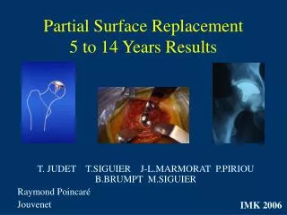
Partial Surface Replacement 5 to 14 Years Results. T. JUDET T.SIGUIER J-L.MARMORAT P.PIRIOU B.BRUMPT M.SIGUIER Raymond Poincaré Jouvenet. IMK 2006. Osteonecrosis mainly affects young patients: How to avoid THR?. Core decompression Cancellous bone grafting

E N D
Partial Surface Replacement5 to 14 Years Results T. JUDET T.SIGUIER J-L.MARMORAT P.PIRIOU B.BRUMPT M.SIGUIER Raymond Poincaré Jouvenet IMK 2006
Osteonecrosis mainly affects young patients: How to avoid THR? • Core decompression • Cancellous bone grafting • Vascularized bone grafting • Intertrochanteric osteotomies • Rotationnal basi-cervical ostéotomies
Drawbacks, limits and failures of conservative surgery • Technically over-demanding procedure • Long lasting rehabilitation • Unpredictable results • Controversial efficiency • Major anatomical disturbance of the upper femur
Partial resurfacing conceptMarc Siguier 1990 • Minimalist design and operative technic • Replacement anatomically limited to the pathologic area • Preservation of the hip anatomy • Preservation of the mecanical properties of the femoral neck
The MMS Implant • 120° Covering area • Range 40 to 60 mm diameter to match the exact sphericity (2mm increment) • Dedicated instrumentation for each size • Cemented fixation
Surgical technic • Anterior approach on fracture table • T-shaped capsulotomy • Anterior dislocation preserving circumflex vessels • Excision of the ostéochondral collapse and under-lying loose necrotic bone
Surgical technic • Measurement of the femoral head • Preparation of the prosthesis setting in line with the center of the head
Surgical technic • Trial implantation without tilting and flush or beneath the surface of the healthy remaining cartilage • Cementation of the definitive prosthesis
Post operative care Immediate rehabilitation and full weight-bearing
Interrogations and potential problems • Quality of bone-implant fixation • Evolutivity of the pre-OP necrotic area • Post-OP extension of the necrotic area toward the pre OP limits • Cartilage tolerance to the implant and further degenerative changes
Previous published results • Siguier M , Judet T, Siguier T & al J.Arthroplasty 1999 Preliminary results of partial surface replacement of the femoral head in osteonecrosis 25 procedures 6 failures (FU 20-60 mths) • Siguier T, Siguier M, Judet T & al Clin. Orthop. 2001 Partial resurfacing arthroplasty of the femoral head in avascular necrosis Methode, indication and results 37 procedures 9 failures (FU 24-89 mths)
Actual Serie • Continuous Serie April 1991 - Jan 2001 • 61 MMS procedures (54 patients) • Age 42 years (24 - 59) • 45 Males et 9 females • Aetiologies Post trauma 6 cases Steroïds 11 Cases (9 patients) Alcool/tobacco 7 cases (5 patients) Idiopathic 35 cases (32 patients)
Clinical Pre- operative Status • Postel Merle-d’Aubigné score 12 pts (8 - 17) Pain 2,3 pts (1 - 4) No preventive surgery
Radiological Pre-operative status • Ficat & Arlet Staging • Stade II : 1 case • Stade III : 41 cases (67 %) • Stade IV : 19 cases (31 %)
Radiological Pre-operative status • Evaluation of the necrotic area size after M.Kerboul -Angular measurement on AP view and Lequesne false profile view Mean angle AP : 121° (70 to 180) Mean angle Profile : 116° (50 to 180)
Radiological Pre-operative status • Evaluation of the necrotic area size -Max depth of the necrotic area; 4 zones to define superficial or deep involvement Stage 2 : 28 % Stage 3 : 53 % Stage 4 : 19 %
Results : 56 over 61 implants1 deceased (2 hips) and2 patients (3 hips) lost to FU before 5 Yrs • Implant removal : 31 cases • Implant still in place : 25 cases
Results : Implant removal : 31 cases • early technical failure • extension of the necrotic zone - head collapse
Results : Implant removal : 31 cases • sinking of the implant • joint line narrowing and degenerative changes No difference statistically significant : failure vs Ficat staging Difference statistically significant : failure vs depth (p < 0,01)
Survival curve • End point : implant removal
Second look surgery • THR through the same mini invasive anterior approach • Resurfacing arthroplasty • No difficulty related to the previous surgery
Conclusion • Optimistic : for 25 patients 40 yrs old, improvement lasting 8 years (5 - 14,5) with a pain scoring rising from 2,3 to 5,1. No deleterious effect on secondary procedure • Realistic : survey curve 50 % à 10 years 46,5 % à 14 years
Conclusion • Prospective in the up to date context of mini-invasive surgery, mini-implants are highly attractive and our long term experience of partial hemi resurfacing of the femoral head must render any surgeon very prudent concerning analogous procedures.