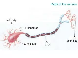
Cell body
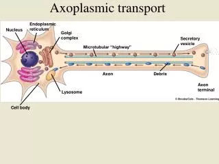
Axoplasmic transport. Endoplasmic reticulum. Nucleus. Golgi complex. Secretory vesicle. Microtubular “highway”. Axon. Debris. Axon terminal. Lysosome. Cell body. Neurocytology & Tract-tracing. Widely used techniques for studying neurons and circuits:

Cell body
E N D
Presentation Transcript
Axoplasmic transport Endoplasmic reticulum Nucleus Golgi complex Secretory vesicle Microtubular “highway” Axon Debris Axon terminal Lysosome Cell body
Neurocytology& Tract-tracing Widely used techniques for studying neurons and circuits: Visualization of neurons Nissl staining, Golgi methods, intracellular dye injections, immunohistochemistry Degeneration and reactive changes in the neuron after lesion Wallerian degeneration Axonal transport methods Autoradiography, HRP, Lectins, Biocytin, Dextrans, Fluorescent Tracers
The Golgi method cerebellar Purkinje cell
Intracellular injectionof Lucifer Yellow Biolistics (“gene-gun”)
Immunohistochemistry L7 protein reveals cerebellar Purkinje cells PEP-19 antiserum reveals the calyx of Held
Tract-Tracing Retrograde degeneration Anterograde Wallerian degeneration Anterograde Degeneration: Reduced silver method and electron microscopy
Collateral projections Labeled terminals Anterograde transport Uptake by Cell body AnterogradeTract-tracing Autoradiography Radioactively labeled amino acid
RetrogradeTract-Tracing HRP, Dextran Retrograde transport Uptake by terminals HRP
Protection of the CNS • The cranium encloses the brain, and the vertebral column encloses the spinal cord. • The CNS is wrapped by several meninges: the outer dura mater, the middle arachnoid mater, and the innermost pia mater. • The brain is surrounded by (and suspended in) the cerebrospinal fluid (CSF). • The blood-brain barrier limits access of blood-borne substances to the brain.
Cranial cavity: contents • The cranial cavity (and vertebral canal) are closed, relatively isolated spaces. • Basic boundary is the arachnoid mater • Contents include: • Brain and spinal cord (intra- and extracellular fluid) • CSF • Blood CSF formation = ~500 ml/day brain = 1500 ml (1200 ICF; 300 ECF) CSF = 125 ml (30 in ventricles) blood = 80 ml dura arachnoid
Right lateral ventricle Left lateral ventricle Third ventricle Central canal of spinal cord Fourth ventricle
The CSF is formed and circulates in the ventricles. • It is produced by the choroid plexuses inside the ventricles, and circulates through the ventricles. • From the fourth ventricle it enters the subarachnoid space, between the arachnoid mater and pia mater. • Arachnoid villi in this space drain the CSF into the blood.
Scalp Skull bone Dura mater Dural sinus Arachnoid villus Arachnoid mater Subarachnoid space of brain Pia mater Venous sinus Brain (cerebrum)
CSF composition CSF production • clear and colorless • little/no protein • acellular (0-5 wbc/ml is normal) • low Glucose (30% below plasma) • Ions: = Na+, Cl, K+, Ca++ (comp. to plasma) • pH =7.33 • *cloudy, colored, cellular CSF implies pathology • >80% from choroid plexus • (specialized ependymal cells) • rate = .3-.4 ml/min • (~500ml total vol./day) • pressure=<200 mm H2O
CSF pathology • too much anywhere = hydrocephalus • excess production = quite rare • impeded circulation = “non-communicating” hc (blockages) • impeded drainage = “communicating” hc due to failed reabsorption • leaking from head = skull fracture • altered composition = bleeds, infections, tumors
Cerebral circulation • Brain = 3 - 4% of body weight • gets 15-18% of cardiac output • uses 20% of total O2 consumed • specialized barrier functions (BBB) • specific areas which lack BBB
Space containing cerebrospinal fluid Ependymal cell Neurons Astrocyte Capillary Microglial cell Oligodendrocyte
Functional blood-brain barrier • allows: small lipophilic molecules; substances with mediated transport (amino acids, glucose) • blocks: large, charged, hydrophilic molecules; some therapeutics (antibiotics) • imaging can detect “leaks” indicating pathology
Top Corpus callosum Cerebral cortex Front of brain Thalamus (wall of third ventricular cavity) Pineal gland Hypothalamus Cerebellum Pituitary gland Brain stem Spinal cord
The CNS consists of the brain and spinal cord. • The outline for brain anatomy is: • Brain stem • Cerebellum • Forebrain • Diencephalon • Hypothalamus • Thalamus • Cerebrum • Basal nuclei • Cerebral cortex
Table 5.3 (1)Page 144 Brain component Cerebral cortex Cerebral cortex Basal nuclei (lateral to thalamus) Basal nuclei Thalamus (medial) Thalamus Hypothalamus Hypothalamus Cerebellum Cerebellum Midbrain Brain stem (midbrain, pons, and medulla) Brain stem Pons Medulla Spinal cord
Major Functions 1. Sensory perception 2. Voluntary control of movement 3. Language 4. Personality traits 5. Sophisticated mental events, such as thinking memory, decision making, creativity, and self-consciousness 1. Inhibition of muscle tone 2. Coordination of slow, sustained movements 3. Suppression of useless patterns of movements 1. Relay station for all synaptic input 2. Crude awareness of sensation 3. Some degree of consciousness 4. Role in motor control 1. Regulation of many homeostatic functions, such as temperature control, thirst, urine output, and food intake 2. Important link between nervous and endocrine systems 3. Extensive involvement with emotion and basic behavioral patterns 1. Maintenance of balance 2. Enhancement of muscle tone 3. Coordination and planning of skilled voluntary muscle activity 1. Origin of majority of peripheral cranial nerves 2. Cardiovascular, respiratory, and digestive control centers 3. Regulation of muscle reflexes involved with equilibrium and posture 4. Reception and integration of all synaptic input from spinal cord; arousal and activation of cerebral cortex 5. Role in sleep-wake cycle
The basal nuclei have an inhibitory role in motor control: • inhibiting muscle tone throughout the body • selecting and maintaining purposeful muscle activity while inhibiting useless movement • monitoring and controlling slow, sustained contractions • Implicated in Parkinson’s Disease (dopamine deficiency) • Increased muscle tone; resting tremors; slow initiation of movement
The limbic system Frontal lobe • functions with the higher cortex. • plays a key role in emotion. • works with the higher cerebral cortex to control behavioral patterns. • the limbic system has reward and punishment centers. • neurotransmitters in the pathways for emotional behavior include norepinephrine, dopamine, and serotonin. Cingulate gyrus Fornix Thalamus Hippocampus Temporal lobe Amygdala Hypothalamus Olfactory bulb
Median sagittal section of cerebellum and brain stem Regulation of muscle tone, coordination of skilled voluntary movement Planning and initiation of voluntary activity Maintenance of balance, control of eye movements Vestibulocerebellum Spinocerebellum Cerebrocerebelum
Motor cortex Informed of motor command Spinocerebellum Motor command to muscles Makes adjustments as necessary Informed of actual performance Activates receptors in muscles and joints Skeletal muscles Movement
The cerebral cortex has four lobes, each is specialized for different activities. • The lobes and some of their functions: • Occipital lobe- initial processing of visual input • Temporal lobe - integration of multiple sensory inputs, primary auditory cortex, Wernicke’s area • Parietal lobe - somatosensory processing. Each region of parietal cortex receives somesthetic and proprioceptive input from a specific body area, mostly from the opposite side of the body. • Frontal lobe - voluntary motor activity, speaking ability (Broca’s area), and elaboration of thought. Stimulation of different areas of its primary motor cortex moves different body regions.
Central sulcus Frontal lobe Parietal lobe Parietooccipital notch Occipital lobe Lateral fissure Cerebellum Temporal lobe Brain stem
Occipital lobe Primary visual cortex
Wernicke’s area Primary auditory cortex Temporal lobe
Somatosensory cortex Central sulcus Posterior parietal cortex Parietal lobe Wernicke’s area
Figure 5.11 (2)Page 149 Sensory homunculus Left hemisphere Cross-sectional view Temporal lobe
Primary motor cortex Central sulcus Frontal lobe Broca’s area
Motor homunculus Left hemisphere Cross-sectional view Temporal lobe
Other cortices and motor function • supplementary motor cortex - medial surface of hemisphere anterior to primary motor cortex. • Preparatory role in programming complex sequences of movements. Stimulation results in complex movement patterns. Lesions do not result in paralysis, but interfere with integration. • premotor cortex - lateral surface of hemisphere anterior to primary motor cortex. • Orienting body and arms toward specific targets. Must be informed of body’s current position in relation to target. This information is relayed by the posterior parietal cortex. • posterior parietal cortex - posterior to the primary somatosensory cortex. • Integration of somatosensory and visual input- important for complex movements.
Supplementary motor area Primary motor cortex Central sulcus Premotor cortex Posterior parietal cortex Frontal lobe
Association areas of the cortex carry out many higher functions: • prefrontal association cortex - functions include planning for voluntary activity, decision-making, creativity, and developing personality traits. • parietal-temporal-occipital association cortex - integrates somatic, auditory, and visual sensations from these three lobes. • limbic association cortex - involved with motivation, emotion, and memory
Central sulcus Prefrontal association cortex Parietal-temporal-occipital association cortex Limbic association cortex
Sensory input Primary sensory areas (somatosensory, 1o visual, 1o auditory cortices) Higher sensory areas Association areas Higher motor areas Primary motor areas Motor output
Hemispheric specialization • The left cerebral hemisphere excels in performing logical, analytical, sequential, and language/verbal tasks • The right cerebral hemisphere excels in spatial perception and artistic and musical talents.
Different aspects of language are controlled by different cortical areas. • Broca’s area is responsible for speaking ability. • Wernicke’s area functions for language comprehension. • Various language disorders are localized in different regions of the cerebral cortex. Damage to these areas can explain the origin of these disorders.
Facial area of motor cortex Angular gyrus of parietal-temporal-occipital association cortex Broca’s area Wernicke’s area Bundle of interconnecting fibers Visual cortex