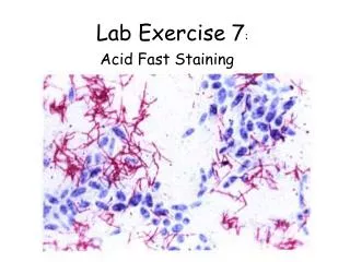

Lab Exercise 7
E N D
Presentation Transcript
Lab Exercise 7 The Integumentary System Portland Community College BI 231
Skin • Or Cutaneous membrane is an organ. • (an organ is a structure that contains two or more of the primary tissue types) • Embedded within the skin are various accessory structures the combination of both is called the integumentary system
Skin • Epidermis: Superficial layer • Made of stratified squamous keratinized epithelium • 4-5 Layers • Dermis: Underlying connective tissue layer • 2 Layers • Hypodermis: Not part of the skin, it is deep to the dermis • Primarily adipose tissue
Layers of the epidermis Stratum basale: single row of cells • Adjacent to the dermis and attached by the basement membrane. • Constantly dividing and pushing up layers • Melanocyteswith melanin protects the skin from UV radiation
Layers of the Epidermis Stratum spinosum: several cell layers superficial to stratum basale. • Cells also dividing • Cells contain bundles of intermediate filaments made of pre-keratin • Cells appear star-shaped because the cell membrane pulls away from the other cells, except in areas where desmosomes are.
Layers of the Epidermis Stratum granulosum: • The upper part of this layer has cells that are beginning to die • Lamellated granules contain a waterproofing glycolipid that is secreted into the extracellular space • Keratohyaline granules combine with intermediate filaments to form keratin fibrils
Layers of the Epidermis Stratum lucidum: Thin translucent layer of dead keratinocytes • Found only in thick skin
Layers of the Epidermis • Stratum corneum: Outermost layer • 20-30 cell layers thick • Cells are dead and flattened • Full of keratin • Constantly being rubbed off
Epidermis cell phases • Deepest layer (basale) goes through rapid division. • Middle layers undergo a process of producing precursor molecules that lead to waterproofing of the cells. • The final phase is the completion of the waterproofing process. Cells eventually die, providing a tough barrier
Dermis • The dermis is the connective tissue layer under the epidermis. • Composed of primarily of fibers: • Irregularly arranged collagen fibers make up the majority and provide strength and flexibility to skin • Lesser numbers of elastic fibers which provided elasticity to the skin • There are also reticular fibers found in the dermis • blood vessels, nerves, sensory receptors, hair follicles and glands.
Dermis • Papillary Layer: Superficial dermal region • Areolar connective tissue • Contains capillaries, lymphatics and sensory neurons • Dermal Papillae: the fingerlike projections from the superior surface • Epidermal ridge: The epidermal layer that dips down into the dermal papillae • Create fingerprints
Epidermis Epidermal ridge PapillaryLayer Dermal Papillae ReticularLayer
Finger Prints Epidermal Ridge
Reticular Layer: Deepest skin layer Dense irregular connective tissue Contains the arteries, veins, sweat and sebaceous glands Cleavage lines: the deep creases (like in the palm) where collagen and elastic fibers are arranged in parallel bundles. Dermis
Hypodermis Dermis • Subcutaneous layer that is not part of the skin • Beneath the dermis layer • Composed of adipose and areolar connective tissue • Highly vascular Hypodermis
Cells of the Epidermis • Keratinocytes: The main cells of the epidermis • Produce keratin, water proof protein • As new cells form, they push the older cells toward the surface, where they gradually accumulate keratin and eventually die • Melanocytes: Spidery black cells • In stratum basale • Produce melanin, the pigment that protects skin from UV damage
Meissner’s (Tactile) Corpuscle • Located in the dermal papillae • Receptor for light touch
Merkel Cells • Merkel Cells: At the junction of the sensory nerve endings • In upper dermis and ll lower epidermis Light touch (like fly walking over cheek)
Pacinian (Lamellated) Corpuscle • Lie deep in the dermis • Respond only when deep pressure is first applied • Monitor high frequency vibrations
Other skin receptors • Warm and cool receptors: When you are at comportable temperature both of these are firing • If you increase or decrease the skin temperature beyond the range of these receptors, pain receptors are stimulated. • Pain receptors are naked nerve endings in the dermis that respond to numerous environmental stimuli.
Integumentary Glands • Sudoriferous (sweat) glands • Lactiferous (milk) glands • Sebaceous (oil) glands • Ceruminous (earwax) glands
Eccrine (Merocrine) Sweat Glands • Eccrine sweat glands • Ducts open directly on the surface of the epidermis • Produce normal body perspiration. • Reduces body temperature by evaporative cooling
Apocrine Sweat Glands • Apocrine glands: secrete a water and a higher concentration of organic acids than eccrine glands that bacteria can use for nutrients (creates body odor) • Found in the armpits, around nipples and in the pubic region • Secrete products into hair follicles or directly onto the surface. • Begin functioning at puberty
Apocrine Sweat Glands • Red arrow - Apocrine Sweat Glands • Green arrow - Hair follicle
Sebaceous Glands • Sebaceous glands • Produce oily substance called sebum • Helps waterproof the skin • Acne: infection of the sebaceous gland
Keratinized cells produced in Hair follicles: tubular compartments that contain hair(projections of the epidermis into dermis) Hair
Hair • Bulb: deepest portion of the hair follicle • Bulb contains the hair matrix which contains actively dividing cells • The actively dividing cells give rise to the hair root. As hair root approaches the surface of the skin it becomes the hair shaft. Hard Keratin covers the hair
Arrector Pili Muscle • Arrector pili muscle: Smooth muscle that pull hair upright during fright or cold (goose bumps)
Hair • Determinate hair grows to a specific length and then stops • Found in axilla, groin, eyelashes and eyebrows. • Indeterminate hair grows without regard to length. • Found on scalp and beards
Hair • Medulla central portion of the hair which is enclosed by an outer cortex. • The cortex may contain pigments which give the hair its color (melanin) • The cuticle is superficial to the cortex
Fingernails • Scale like modification of the epidermis • Free edge: part that grows away from the finger • Body: The nail • Root: Embedded in skin and sticks to the nail bed • Nail Bed: Extension of the stratum basale beneath the nail • Nail Matrix: Proximal part of the nail bed responsible for nail growth • Lunula: white crescent area; Most active growth region of nail matrix
Muscle Tissue • Like epithelial tissue, muscular tissue is a cellular tissue with the tissue having mostly cells and little matrix • There are three types of muscle: skeletal, cardiac and smooth
Skeletal Muscle • Cells called fibers • Striated • Voluntary • Multinucleate
Cardiac Muscle • Striated but striations are much less obvious. • Cardiac muscle cells are called myocytes. • Branched • Involuntary • Intercalated discs, which facilitate the transmission of the electrical impulses in the heart
Smooth Muscle • Nonstriated • Involuntary • Cells of smooth muscle are spindle-shaped • Found in intestine where it propels food along by a process known as peristalsis and segmentation
The End The End