Pulmonary Structure & Function
Explore the anatomy of the ventilatory system, lung structures, ventilation mechanics, and gas exchange processes to grasp the complexities and functions of the pulmonary system.

Pulmonary Structure & Function
E N D
Presentation Transcript
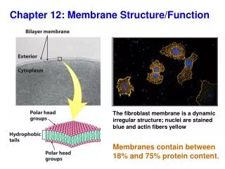
Chapter 12 Pulmonary Structure and Function
Chapter Objectives • Diagram and labelthe main structures of the ventilatory system components • Describe the ventilatory system’s conducting, transitional, and respiratory zones • Discuss the mechanical and muscular aspects of inspiration and expiration during rest and physical activity • Define and quantify static and dynamic lung function measures at rest and exercise
Chapter Objectives, cont. • Describe two effects of cold-weather exercise on the respiratory tract • Discuss the contributions of breathing rate and tidal volume to minute ventilation and alveolar minute ventilation at rest and physical activity • Discuss two factors that account for variations in the ventilation–perfusion ratio among healthy individuals
Chapter Objectives, cont. • Explain the four phases of the Valsalva and discuss the physiologic consequences of this maneuver • Define minute ventilation, alveolar ventilation, ventilation perfusion ratio, and anatomic and physiologic dead space
Anatomy of Ventilation • Pulmonary ventilation moves and exchanges ambient air with air in the lungs • Ambient air enters the nose and mouth trachea (and adjusts to body temperature), is filtered and humidified two bronchi bronchioles alveoli
The Lungs • Provide the gas exchange surface that separates blood from surrounding gaseous environment • O2 transfers from alveolar air into alveolar capillary blood while the blood’s CO2 moves into the alveolar the alveoli and then into ambient air • An average-sized adult’slung weighs about 1 kg with a volume of 4 to 6 L • The lungs consists of 10% solid tissue, with the remainder filled with air and blood
The Alveoli • More than 600 million alveoli provide the surface for gas exchange between lung tissue and blood • Alveoli receivethe largest blood supply of all organs • Capillaries and alveoli lie side-by-side with thin surfaces to facilitate rapid gas exchange • Pores of Kohn within alveoli evenly disperse surfactant over respiratory membranes to reduce surface tension for easier alveolar inflation • Each minute at rest, 250 mL O2 leave the alveoli and enter the blood and 200 mL CO2 diffuse in the opposite direction
Zones of Ventilation • Ventilatory subdivisions • Conducting zones: Trachea and terminal bronchioles • Considered anatomic dead space • Function in air transport, humidification, warming, particle filtration, vocalization, immunoglobulin secretion • Transitional and respiratory zones: Bronchioles, alveolar ducts, and alveoli • Functionin gas exchange, surfactant production, molecule activation and inactivation, blood clotting regulation, and endocrine function
Zones of Ventilation,cont. • More than 600 million alveoli provide the surface for gas exchange between lung tissue and blood
Lung Airflow Related to Total Cross-sectional Tissue Area • Forward airflow velocity during inspiration decreases because of the large increase in tissue cross-sectional area in the terminal bronchiole region
Fick’s Law of Diffusion • Governs gas diffusion across a fluid membrane • Gas diffuses through a sheet of tissue at a rate: • directly proportional to tissue area, a diffusion constant, and pressure differential of the gas on each side of the membrane • inversely proportional to tissue thickness • The pressure differential between air in the lungs and lung–chest wall interface causes lungs to adhere to the chest wall and follow its every movement
Inspiration • Phases: • Diaphragm contracts, flattens, and moves downward toward the abdominal cavity • Elongation and enlargement of the chest cavity expands air in the lungs, causing its intrapulmonic pressure to decrease slightlybelow atmospheric pressure • Lungs inflate as nose and mouth suck air inward • Completed when thoracic cavity expansion ceases; this causes equality between intrapulmonic and ambient atmospheric pressures
Expiration • The passive process during rest and light exercise as air moves out of lungs from: • Natural recoil of stretched lung tissue and relaxation of inspiratory muscles • Expiration Phases • Sternum and ribs drop, diaphragm rises (decreasing chest cavity volume and compressing alveolar gas), moving air from respiratory tract to the environment • Completes when compressive force of expiratory muscles (activated in vigorous exercise) cease and intrapulmonic pressure decreases to atmospheric pressure
Surfactant (Surface Active Agent) • “Wetting agent” lipoprotein mixture of proteins, phospholipids, and calcium ions produced by alveolar epithelial cells (to reduce surface tension) • Mixes with fluids encircling alveolar chambers • Interrupts the surrounding water layer, reducing the alveolar membrane’s surface tension, thereby increasing overall lung compliance • Reduces the energy required for alveolar inflation and deflation • Without surfactant, small alveoli would collapse (atelectasis ) from high collapsing pressures
Lung Volumes and Capacities: Static Lung Volumes • Tidal volume: Air moved during the inspiratory or expiratory phase of each breathing cycle (0.4 to 1.0 L of air per breath) • Inspiratory reserve volume: Inspiring as deeply as possible following a normal inspiration (2.5 to 3.5 L above inspired tidal air) • Expiratory reserve volume: After a normal exhalation, continuing to exhale and forcing as much air as possible from the lungs (1.0 to 1.5 L) • Forced vital capacity: Total volume of air voluntarily moved in one maximalbreath; it includes TV plus IRV and ERV (4 to 5 L in young men) and (3 to 4 L) in young women
Residual Lung Volume • Air volume remaining in the lungs after a forced maximal exhalation • Averages 0.8 to 1.2 L forhealthy college-aged women, and 0.9 to 1.4 L for college-aged men • Increases with age • Allows an uninterrupted exchange of gas between the blood and alveoli to prevent fluctuations in blood gases during phases of the breathing cycle • RLV plus FVC = total lung capacity (TLC)
Effects of Previous Exercise Effects on RLV • RLV temporarily increases from an acute bout of either short-term or prolonged exercise due to: • Closure of small peripheral airways • Increase in thoracic blood volume
Dynamic Lung Volumes • Dynamic pulmonary ventilation depends on: • Maximum “stroke volume” of the lungs (FVC) • Speed of moving a volume of air (breathing rate) • Determined by lung compliance (the resistance of respiratory passages to air movement and “stiffness” imposed by the chest and lung tissues)
FEV-to-FVC Ratio • Forced expiratory volume (FEV) • Maximal airflow measured over 1 s (FEV1.0) • FEV1.0 ÷ FVC indicates pulmonary airflow capacity • Reflects pulmonary expiratory power and overall resistance to air movement upstream in lungs • FEV1.0 ÷ FVC = ~85% in healthy individuals • FEV1.0 ÷ FVC ≤70% = delineation point for airway obstruction
Examples of Pulmonary Function Tests in Normal Subjects and in Patients with Obstructive or Restrictive Lung Disease
Maximum Voluntary Ventilation (MVV) • Evaluates ventilatory capacity with rapid and deep breathing for 15s • Extrapolated to volume if continued for 1 min • MVV typically ranges between 35 to 40 times FEV1.0 • MVV ranges between 140 and 180 L/min in healthy, college-age men, and 80 and 120 L/min in healthy, college-age women • Exercise training of the ventilatory muscles improves their strength and endurance, increasing inspiratory muscle function and MVV
Physical Activity Implications of Gender Differences in Static and Dynamic Lung Function Measures • Compared to men, women have a reduced lung size and airway diameter, a smaller diffusion surface, and lower static and dynamic lung function measures • Leads to expiratory flow limitations, greater respiratory muscle work and use of ventilatory reserve during maximal exercise (particularly in highly trained women) • A smaller lung volume plus a high expiratory flow rate demands in trained women during intense exercise places considerable demand on maximum flow–volume envelope of the airways; this adversely affects the maintenance of alveolar-to-arterial oxygen exchange
Lung Function, Aerobic Fitness, and Physical Performance • Regular endurance exercise does not stimulate large increases in pulmonary system functional capacity • Dynamic lung function tests indicate severity of obstructive and restrictivelung diseases, but provide little information about aerobic fitness or exercise performance when values fall within the normal range
Pulmonary Ventilation • Can be viewed from two perspectives: • Volume of air moved into or out of the respiratory tract each minute • Air volume that ventilates only alveolar chambers each minute
Minute Ventilation (VE) • The volume of air breathed each minute VE = Breathing rate × Tidal volume • can be increased by an increase in breathing rate or breathing depth, or both • breathing rate can increase to 35 to 45 breaths/min during strenuous exercise in healthy young adults and 60 to 70 breaths/min in some elite endurance athletes • Tidal volume for trained and untrained individuals rarely exceed 60% of vital capacity
Alveolar Ventilation • Anatomic dead space: air in each breath that does not enter alveoli and participate in gaseous exchange with blood (range 150 to 200 mL) • Alveolar ventilation:portion of inspired air reaching the alveoli and participating in gas exchange • Determines the gaseous concentrations at the alveolar–capillary membrane • About 350 mL of the 500 mL of inspired TV at rest enters into and mixes with existing alveolar air
Typical Values for Pulmonary Ventilation During Rest and Physical Activity
Dead Space Versus Tidal Volume • Anatomic dead space increases as TV becomes larger; it often doubles during deep breathing from stretching of respiratory passages with a fuller inspiration • Any increase in dead space represents proportionately less volume than the increase in TV • Deeper breathing provides more effective alveolar ventilation than similar minute ventilation achieved through increased breathing rate
Ventilation-Perfusion (V-P) Ratio • The ratio of alveolar ventilation to pulmonary blood flow • ~4.2 L of air ventilates alveoli each min at rest and ~5.0 L of blood flows through pulmonary capillaries each min • Average V-P Ratio = 0.84 - An alveolar ventilation of 0.84 L matches each liter of pulmonary blood flow • In light exercise, V-P Ratio remains ~0.8 L • In intense exercise V-P Ratio = 5.0L
Physiologic Dead Space • The portion of the alveolar volume with a ventilation–perfusion ratio that approaches zero • Sometimes alveoli may not function adequately in gas exchange because of two factors: • Underperfusion of blood • Inadequate ventilation relative to alveolar surface • In certain pathologic conditions, physiologic dead space increases to 50% of TV • Adequate gas exchange becomes impossible when dead space of the lung exceeds 60% TLV
Distribution of Tidal Volume • TV includes about 350 mL of ambient air mixed with alveolar air, 150 mL ambient air remaining in air passages (anatomic dead space), and a small portion of air in the physiologic dead space
Breathing Rate Versus Tidal Volume • Increasing the rate and depth of breathing increases alveolar ventilation in physical activity • In moderate activity, well-trained athletes maintain alveolar ventilation by increasing TV with only a small increase in breathing rate • In exercise, breathing becomes deeper and alveolar ventilation increases from 70% at rest to >85% of exercise minute ventilation • Each person develops their “style” of breathing; breathing rate and TV blend to provide effective alveolar ventilation
Variations from Normal Breathing Patterns • Hyperventilation • Increase in pulmonary ventilation that exceeds the O2 consumption and CO2 elimination needs of metabolism • Dyspnea • Inordinate shortness of breath or subjective breathing distress • Valsalva Maneuver • Closing glottis following a full inspiration while maximally activating expiratory muscles, creating compressive forces that increase intrathoracic pressure above atmospheric pressure • Occurs commonly in activities that require a rapid, maximum application of force forshort duration
Physiologic Consequences of Performing the Valsalva Maneuver • Performing a prolonged Valsalva maneuver during static, straining-type exercise dramatically reduces venous return and arterial blood pressure • Diminishes brain’s blood supply, producing dizziness or fainting • Once the glottis reopens and intrathoracic pressure normalizes, blood flow reestablishes with an “overshoot” in arterial blood pressure • Does not cause the large increases in blood pressure during heavy resistance exercises; these exercises greatly increase resistance to blood flow in active muscle with a resulting rise in systemic blood pressure
Valsalva Maneuver • Valsalva reduces return of blood to the heart because increased intrathoracic pressure collapses inferior vena cava that runs through the chest cavity Straining exercise with Valsalva Normal breathing
Valsalva Maneuver, cont. • Valsalva reduces return of blood to the heart because increased intrathoracic pressure collapses inferior vena cava that runs through the chest cavity Normal aortic pulse pressure with Valsalva during calibrated muscle strain
Respiratory Tract During Cold-Weather Activity • Cold ambient air normally does not damage respiratory passages because of relatively rapid airway warming • This air-conditioning process greatly increases air’s capacity to hold moisture, which produces measurable water loss from the respiratory passages • In cold weather, the respiratory tract loses considerable water and heat, especially during strenuous exercise • Fluid loss from the airways contributes to dehydration, dry mouth, burning sensation in the throat, and generalized irritation of the respiratory passages
Summary • The lungs provide a large surface between the body’s internal fluid environment and the gaseous external environment. During any 1 s of physical activity, no more than 1 pint of blood flows in the pulmonary capillaries. • Fick’s law of diffusion governs gas movement across a fluid membrane • Surfactant consists of a lipoprotein mixture secreted within lung tissue that reduces surface tension between the alveolar membrane and surrounding tissues
Summary, cont. • Pulmonary airflow depends on small pressure differentials between ambient air and air within the lungs. Ventilatory muscle action produces these pressure differences • Lung volumes vary with age, gender, and body size (particularly stature) • The residual lung volume represents air remaining in the lungs following maximal exhalation • FEV and MVV dynamically measure the ability to sustain a high airflow level
Summary, cont. • Measures of static and dynamic lung function within the normal range poorly predict aerobic fitness and exercise performance • Breathing rate and tidal volume (TV) determine pulmonary minute ventilation • Alveolar ventilation reflects the portion of minute ventilation that enters the alveoli for gaseous exchange with the blood • The ventilation–perfusion ratio reflects the association between alveolar minute ventilation and pulmonary blood flow
Summary, cont. • TV increases during physical activity by encroachment into inspiratory and expiratory reserve volumes • During intense exercise, TV plateaus at approximately 60% of the vital capacity; minute ventilation increases further through increases in breathing rate
Summary, cont. • A Valsalva maneuver describes a forced exhalation against a closed glottis, which causes large pressure increases within the chest and abdominal cavities that compress the thoracic veins (thereby reducing venous return to the heart) • Breathing cold ambient air normally does not damage respiratory passage
Image Sources Slide 12: Adapted with permission from West JB. Respiratory Physiology—The Essentials. 8th ed. Baltimore: Lippincott Williams & Wilkins, 2008. Slide 40: Data from Hébert J-L, et al. Pulse pressure response to the strain of the Valsalva maneuver in humans with preserved systolic function. J Appl Physiol 1998;85:817.