Pulmonary Function Testing
Pulmonary Function Testing. Let’s catch our breath Eddie Needham, MD, FAAFP Program Director Emory Family Medicine Residency Program. Learning Objectives The Astute Learner will:. Become familiar with indications for performing PFTs. Become adept at interpreting PFTs.

Pulmonary Function Testing
E N D
Presentation Transcript
Pulmonary Function Testing Let’s catch our breath Eddie Needham, MD, FAAFP Program Director Emory Family Medicine Residency Program
Learning ObjectivesThe Astute Learner will: • Become familiar with indications for performing PFTs. • Become adept at interpreting PFTs. • Perform and interpret a PFT on a colleague. • Breath deep – it feels good
Lung Volumes and Capacities • There are four basic lung volumes: • Inspiratory reserve volume (IRV) • Tidal volume (TV) • Expiratory reserve volume (ERV) • Residual volume (RV) • In various combinations, these lung volumes then form lung capacities. • E.g., Vital capacity = IRV + TV + ERV
Indications for Pulmonary Function Testing • Patients 45 years old and older who have ever smoked. • Patients with prolonged or excessive cough or sputum production. • Patients with a history of exposure to lung irritants. Petty TL et al, NHLBI Workshop Summary, JAMA, 1997; 277: 246-53.
Indications for Pulmonary Function Testing • Detecting pulmonary disease • Pulmonary symptoms – chest pain, orthopnea, cough, phlegm production, dyspnea, wheezing • Physical findings – Chest wall problems, cyanosis, clubbing, decreased breath sounds • Abnormal labs/x-rays – ABG, Chest X-Ray Barreiro TJ and Perillo, I. An Approach to Interpreting Spirometry, AFP, March 1, 2004; 69: 1107-14.
Assessing disease severity and progression Pulmonary disease – COPD, Cystic fibrosis, Interstitial lung disease, Sarcoidosis Cardiac disease – CHF, Congenital heart disease, Pulmonary hypertension Neuromuscular disease – Amyotrophic lateral sclerosis, Guillain-Barre syndrome, Multiple sclerosis, Myasthenia gravis Indications for Pulmonary Function Testing Barreiro TJ and Perillo, I. An Approach to Interpreting Spirometry, AFP, March 1, 2004; 69: 1107-14.
Indications for Pulmonary Function Testing • Pre-operative risk stratification • Thoracic surgery • Cardiac surgery • Organ transplantation • General surgical procedures • Evaluating disability and impairment Barreiro TJ and Perillo, I. An Approach to Interpreting Spirometry, AFP, March 1, 2004; 69: 1107-14.
Needham’s Take onIndications for Pulmonary Function Testing • Possible COPD? Just do it • Convincing a smoker to stop? Just do it • Prolonged cough? Just do it • Abnormal physical exam findings? Just do it Slide sponsored by …
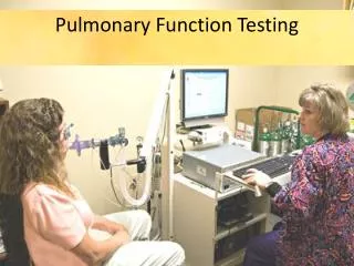
Actual PFT Performance Technique • Prepare the equipment – find a nurse who knows (or is that nose?) what to do. • Patient should be seated with nose clip in place. • The patient needs to practice the exercise before actually performing the test. Have the patient breath in and out deeply several times. • Ask the patient to breath in as deeply as they can. Jackson, E. Pfenniger’s Texbook of Procedures, Chapter 62, 485-492.
Actual PFT Performance Technique • The patient should place their mouth completely over the mouthpiece, not inside it. • Ask the patient to blow out as fast and as quick as they can for at least six seconds. Enthusiatically coach the patient – jump, shout, get down, hoot and holler… “Blow, blow, come on, blow more, you can do it!” Jackson, E. Pfenniger’s Texbook of Procedures, Chapter 62, 485-492.
Actual PFT Performance Technique • Once the patient has blown out as much as they can, ask them to then inhale as deeply as they can. • Repeat the whole test three times. The goal is to get a reproducible result that is consistent. • You may need to repeat the test more than three times in order to obtain an internally valid test. Jackson, E. Pfenniger’s Texbook of Procedures, Chapter 62, 485-492.
Normal Values • FVC is the total amount of air a person can exhale, usually measured in six seconds. • 80 – 120% of predicted is a normal value • 70 – 80% demonstrates mild reduction/restriction • 50 – 70% demonstrates moderate reduction • <50% demonstrates severe reduction • FEV1 is the amount of air a person can exhale in one second. • 80 – 120% of predicted is a normal value
Normal Values • FEV1/FVC ratio is the percentage of FVC that can be expired in one second. • 75 – 80% is normal • 60 – 80% demonstrates mild obstruction • 50 – 60% demonstrates moderate obstruction • <50% demonstrates severe obstruction
Normal Values • FEF25-75 reflects small airway function • >80% is normal • 60 – 80% reflects mild obstruction in the small airways • 40 – 60% reflects moderate obstruction • <40% reflects severe obstruction
PFT Interpretation • Three steps in interpretation • Is the test valid? • Interpret the test • Classify severity of disease if present
Validity • The test is valid is you have good patient effort and the three tests performed are internally consistent. • You may notice a learning curve in that the latter tests are better performed than the former. • Make sure that the tests are maximal effort. You need to be really aggressive in coaching your patient.
PFT Interpretation • Assess FVC, FEV1, and FEV1/FVC ratio. • FVC and FEV1 normal, with a normal FEV1/FVC ratio: • Normal Test …yeah!!! • FVC decreased, FEV1 low or normal, and a normal to high FEV1/FVC ratio: • Restrictive lung disease • FVC normal or low, FEV1 low, and a low FEV1/FVC ratio: • Obstructive lung disease
Actual Predicted % Predicted FVC 4.0 4.5 88 FEV1 3.4 4.2 89 FEV1/FVC 85 82 112 FEF25-75 Normal
Actual Predicted % Predicted FVC 2.0 4.0 50 FEV1 1.8 3.7 47 FEV1/FVC 90 82 112 FEF25-75 Restrictive Pattern
Actual Predicted % Predicted FVC 4.0 4.5 88 FEV1 2.4 4.2 58 FEV1/FVC 60 82 76 FEF25-75 2.2 4.4 50 Obstructive Pattern
Special Techniques • Beta Agonist Challenge • Methacholine Challenge • DLCO
Beta Agonist Challenge • Perform this when there is a suspicion that the obstructive defect may be reversible –> asthma. • Give the patient a beta agonist treatment (two puffs of an albuterol MDI or an albuterol nebulizer) and repeat the PFTs several minutes later. If you notice a 12% or more increase in FEV1, then you have diagnosed reversible airway disease/asthma.
Diffuse capacity of carbon monoxide in the lung DLCO • After performing the standard PFTs, the patient then inhales trace amounts of carbon monoxide. • CO traverses the alveolar capillary beds much more readily than CO2 or O2. • As such, most of the CO inhaled should be absorbed. • When it is not, this suggests pulmonary scarring consistent with pulmonary fibrosis. Search for a cause.
Methacholine Challenge • If you have a suspicion that the patient might have exercise-induced bronchospasm (EIB), then refer them to a pulmonary lab where they can do provocative testing with methacholine. • If the patient has a decrease in their FEV1/FVC ratio with the inhalation of methacholine, then you have diagnosed EIB. • Pretreat before exercise with albuterol or cromolyn.
Case 1 Actual Predicted % Predicted FVC 3.8 4.5 83 FEV1 2.2 4.2 47 FEV1/FVC 59 82 72 FEF25-75 1.6 3.7 43 Survey says … COPD
Case 2 Actual Predicted % Predicted FVC 2.9 4.5 64 FEV1 2.5 4.2 59 FEV1/FVC 89 82 113 FEF25-75 3.7 3.5 102
Case 2 Actual Predicted % Predicted FVC 2.9 4.5 64 FEV1 2.5 4.2 59 FEV1/FVC 89 82 113 FEF25-75 3.7 3.5 102 DLCO is decreased when measured Restrictive lung pattern from Amiodarone
Case 3 Actual Predicted % Predicted FVC 4.0 4.5 88 FEV1 2.6 4.2 57 FEV1/FVC 65 82 71 FEF25-75 1.7 3.6 47 Beta agonist treatment Actual Predicted % Predicted FVC 4.1 4.5 91 FEV1 3.6 4.2 89 FEV1/FVC 90 82 112 FEF25-75 3.2 3.6 91 Reversible obstructive defect, A.K.A ???
Case 4 Actual Predicted % Predicted FVC 4.0 4.5 88 FEV1 3.6 4.2 89 FEV1/FVC 90 82 112 FEF25-75 3.1 3.4 95 Normal
Case 5 Actual Predicted % Predicted FVC 4.0 4.5 88 FEV1 3.3 4.2 81 FEV1/FVC 83 82 101 FEF25-75 1.7 3.5 48 Small Airways Defect
Case 6 Actual Predicted % Predicted FVC 3.5 5.3 68 FEV1 3.1 4.6 68 FEV1/FVC 93 82 117 FEF25-75 3.7 3.3 120 By the way, the patient’s BMI = 47 … Restrictive pattern in obese patient