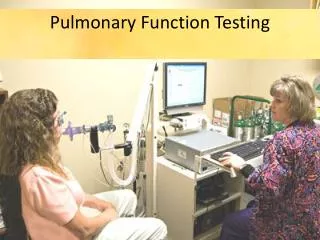
Pulmonary Function Testing
Danish Thameem M.D. Pulmonary and Critical Care Medicine. Pulmonary Function Testing. Indications for Pulmonary Functions. Evaluation of a pulmonary symptom Evaluation of smokers without symptoms Evaluation of workers exposed to hazards Quantification of impairment

Pulmonary Function Testing
E N D
Presentation Transcript
Danish Thameem M.D. Pulmonary and Critical Care Medicine Pulmonary Function Testing
Indications for Pulmonary Functions • Evaluation of a pulmonary symptom • Evaluation of smokers without symptoms • Evaluation of workers exposed to hazards • Quantification of impairment • Evaluate response to therapy • Preoperative assessment • Disability evaluation
Timeline of cigarette smokers that develop obstructive lung disease.
Types of Pulmonary Function Tests • Spirometry • Lung Volumes • Diffusion Capacity • Maximal Respiratory Pressures • Maximum Voluntary Ventilation (MVV) • Arterial Blood Gases • Pulse Oximetry • Bronchoprovocation
Lung Volumes and Capacities • Four Volumes • VT • IRV • ERV • RV • Four Capacities • VC • IC • FRC • TLC
General Approach to Interpretation • Is the test interpretable? • Are the results normal? Or abnormal? • What is the pattern? • What is the severity? • What does this mean for the patient?
Acceptability Criteria for Spirograms • Free from artifacts • Cough or glottis closure during the first second of exhalation • Early termination or cutoff • Variable effort • Leak • Obstructed mouthpiece • Satisfactory exhalation • 6 sec of exhalation and/or a plateau in the volume-time curve or • Reasonable duration or a plateau in the volume-time curve or • The subject cannot or should not continue to exhale
Repeatability Criteria After three acceptable spirograms have been obtained, apply the following tests • Are the two largest FVCs within 0.2 L of each other? • Are the two largest FEV1s within 0.2 L of each other? • If both of these criteria are met, the test session may be concluded. If both of these criteria are not met, continue testing until: Both of the criteria are met with analysis of additional acceptable spirogramsor • A total of eight tests have been performed or • Save a minimum of three best maneuvers
Spirometry • FVC (forced vital capacity): maximum volume of air that can be exhaled during a forced maneuver (after maximal forced inspiration, TLC) • FEV1 (forced expired volume in one second): volume expired in the first second of maximal expiration after a maximal inspiration • FEV1/FVC: FEV1 expressed as a % of FVC, a clinically useful index of airflow limitation
Predicting Normal Values • Depend on patient’s • Height • Age • Gender • Racial & ethnic background • Weight & BMI (to a lesser degree) • Reference Standards
Percent Predicted as Normal Range • Results are expressed as % Predicted of a predicted normal value of a person the same age, sex, and height. (FVC and FEV1) • Normal Ranges • FVC 80-120% • FEV1 80-120% • FEV1/FVC >0.70 of predicted ratio
Obstruction vs. Restriction • If the FVC and / or FEV1 is below normal • The distinction between obstruction & restriction is based on the FEV1/FVC ratio • NIH/WHO - GOLD guidelines recommends using ratio below 0.70 for the diagnosis of COPD
Obstructive Lung Disease • Emphysema & Chronic Bronchitis • Cystic Fibrosis • Asthma • Bronchiectasis • Some Interstitial Lung Disease: (combined)
Restrictive Pattern • Normal or elevated FEV1/FVC ratio • With a low FEV1 or FVC suggests restriction • Lung Volumes are needed to confirm • Some patients with Asthma or COPD may have this pattern (“pseudorestriction”)
Rating of Severity • May be based on statements such as from the American Thoracic Society (ATS) • Obstructive Pattern - FEV1 • Restrictive Pattern – TLC (lung volumes) • If lung volumes not obtained - FVC
ATS/ERS Standardization of Lung Function Testing: Interpretative Strategies for lung function tests - 2005
Classification of COPD by SeverityGOLD Guidelines - 2009 I: Mild FEV1/FVC < 70%; FEV1 > 80% predicted II: ModerateFEV1/FVC < 70%; 50% <FEV1 < 80% III: SevereFEV1/FVC < 70%; 30% < FEV1 <50% IV: Very FEV1/FVC < 70%; FEV1 < 30% predictedSevereor FEV1 < 50% predicted plus chronicrespiratoryfailure
Bronchodilator Response • Must use bronchodilator with rapid onset • Albuterol • Levalbuterol • Increase FEV1 or FVC from baseline • By at least 12% • By at least 200 mL • Both values must be met
Upper Airway Obstruction Patterns • Detect obstructive lesions in the major airways. • Characterizes the lesion: Locationof the lesion: • Intrathoracic • Extrathoracic Behaviorof the lesion in rapid inspiration and expiration: • Fixed • Variable
Variable Extrathoracic Obstruction Vocal cord paralysis Goiter Tumor Levitzky MG Pulmonary Physiology, McGraw Hill 4th ed, 1995, p 50
Variable Intrathoracic Obstruction Tracheomalacia Intratracheal tumor Levitzky MG Pulmonary Physiology, McGraw Hill 4th ed, 1995, p 50
Fixed Obstruction Tracheal stenosis/stricture Bilateral vocal cord paralysis Extrinsic compression Levitzky MG Pulmonary Physiology, McGraw Hill 4th ed, 1995, p 50
Diffusion Capacity • Estimates the transfer of oxygen in the alveolar air to the red blood cell. • Factors that influence the diffusion: 1) Area of the alveolar-capillary membrane (A) 2) Thickness of the membrane (T) 3) Driving pressure 4) Hemoglobin 5) Carboxyhemoglobin
Diffusing Capacity • Single-breath DLCO measures the capacity of the lung to transfer gas • Patient exhales to RV then rapidly inhales gas mixture with minute amount of CO. After, 10 second breath-hold at TLC, the patient rapidly exhales & the exhaled gas is analyzed to measure the amount of CO transferred into the capillary blood during the maneuver
Abnormalities of Diffusing Capacity • Decreased in conditions that disrupt the alveolar-capillary surface for gas transfer • Loss of surface area (resection, fibrosis, emphysema, pneumonia) • Reduced lung capillary volume (vasculitis, thromboembolism, primary pulmhtn, ILD) • Increased diffusion distance (PAP, PCP)
Abnormalities of Diffusing Capacity • Increased by conditions that lead to recruitment of pulmonary vascular bed and increase in capillary blood volume • (exercise, mild CHF, asthma) • Or by increased amount of hemoglobin which binds CO • (pulmonary hemorrhage, erythrocytosis)
CASE 1 • 54 y/o male smoker • PFT • FEV1 : 1.3 L (23%) • FVC : 2.3 L (45%) • FEV1/FVC : 56 • TLC 98% • RV : 156% • DLCO : 30%
Diagnosis • Very severe obstructive defect • Severe reduction in DLCO • High RV • Air trapping COPD
CASE 2 • 35 y/o F with SLE • FEV1 : (56%) • FVC : (45%) • FEV1/FVC 90 • TLC : 48% • RV: 45% • DLCO : 23% • FEV1 increased by 4% (0.1 L) with bronchodilator testing
Diagnosis • Severe restriction without significant response to bronchodilators • Severe reduction in DLCO ILD PULMONARY FIBROSIS
CASE 3 • 45 y/o female with history of allergic rhinitis and dyspnea on exertion • FEV1 - 3.2 (70%) pre, 4.5 (100%) post BD • FVC - 4.9 (70%) pre, 6.0 (85%) post BD • RATIO - 65% pre and 75% post • TLC - 6 L (100%) • DLCO - 100%
Diagnosis • Mild obstruction with significant response to bronchodilators (normal) • Normal lung volumes and DLCO ASTHMA
CASE 4 • 76 y/o male with weight loss and dyspnea • FEV1 - 4 L ( 85%) • FVC - 5.1 L (80%) • RATIO - 78% • TLC - 6 L ( 82%) • DLCO - 88%
Diagnosis • Normal spirometry • Truncated inspiratory limb of the flow volume loop EXTRATHORACIC OBSTRUCTION