Pulmonary Function Testing
Pulmonary Function Testing. INTRODUCTION. http://www.youtube.com/watch?v=WyhOJR8btCs http://www.youtube.com/watch?v=ZIgQGAWJbyY. Introduction. Pulmonary function testing has come into widespread use since the 1970s.

Pulmonary Function Testing
E N D
Presentation Transcript
INTRODUCTION • http://www.youtube.com/watch?v=WyhOJR8btCs • http://www.youtube.com/watch?v=ZIgQGAWJbyY
Introduction • Pulmonary function testing has come into widespread use since the 1970s. • This has been facilitated by several developments.Because of miniaturization and advances in computer technology, microprocessor devices have become portable and automated with fewer moving parts. Testing equipment, patient maneuvers, and testing techniques have become widely standardized throughout the world through the efforts of professional societies. • Widely accepted normative parameters have been established.
Introduction • Pulmonary function testing is a valuable tool for evaluating the respiratory system, representing an important adjunct to the patient history, various lung imaging studies, and invasive testing such as bronchoscopy and open-lung biopsy. • Insight into underlying pathophysiology can often be gained by comparing the measured values for pulmonary function tests obtained on a patient at any particular point with normative values derived from population studies. • The percentage of predicted normal is used to grade the severity of the abnormality. • Practicing clinicians must become familiar with pulmonary function testing because it is often used in clinical medicine for evaluating respiratory symptoms such as dyspnea and cough, for stratifying preoperative risk, and for diagnosing common diseases such as asthma and chronic obstructive pulmonary disease.
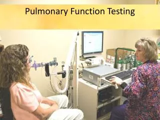
Introduction • Pulmonary function tests (PFTs) is a generic term used to indicate a battery of studies or maneuvers that may be performed using standardized equipment to measure lung function. PFTs can include simple screening spirometry, formal lung volume measurement, diffusing capacity for carbon monoxide, and arterial blood gases. These studies may collectively be referred to as a complete pulmonary function survey.
Introduction • Before a spirogram can be meaningfully interpreted, one needs to inspect the graphic data (the volume-time curve and the flow-volume loop) to ascertain whether the study meets certain well-defined acceptability and reproducibility standards. • Tests that fail to meet these standards can provide useful information about minimum levels of lung function, but, in general, they should be interpreted cautiously. The interpretive strategy usually involves establishing a pattern of abnormality (obstructive, restrictive, or mixed), grading the severity of the abnormality, and assessing trends over time. • Various algorithms are available. Automated spirometry systems usually have built-in software that can generate a preliminary interpretation, especially for spirometry; however, algorithms for other pulmonary function studies are not as well established and necessitate appropriate clinical correlation and physician oversight.
Categories of Pulmonary Function Tests • Pulmonary function studies use a variety of maneuvers to measure and record the properties of four lung components. These include the airways (large and small), lung parenchyma (alveoli, interstitium), pulmonary vasculature, and the bellows-pump mechanism. Various diseases can affect each of these components.
Categories of Pulmonary Function Tests • Measurement of dynamic flow rates of gases through the airways • Measurement of lung volumes and capacities • Measurement of the lung’s ability to diffuse gases
Indications for Pulmonary Function Testing • Evaluation of the cause of pulmonary symptoms such as dyspnea, coughing, wheezing, and exercise intolerance • Evaluation of abnormalities noted on chest X-ray • Determination of the course of a disease and the response to treatment of the disease
Indications for Pulmonary Function Testing • Evaluation of risk for perioperative pulmonary complications • Performance of epidemiological surveillance for pulmonary disease
Indications for Pulmonary Function Testing • Rule out significant pulmonary pathology in people with high risk for pulmonary dysfunction, such as smokers, firefighters, and those exposed to asbestos • Evaluation of degree of disability
Contraindications for Pulmonary Function Testing • Performing lung function tests can be physically demanding for a minority of patients. It is recommended that patients should not be tested within 1 month of a myocardial infarction. • Chest or abdominal pain of any cause • Oral or facial pain exacerbated by a mouthpiece • Stress incontinence • Dementia or confusional state • Patients with any of the conditions are unlikely to achieve optimal or repeatable results • Age of patient, ages of 8 and less render less accurate results
Patient Position for Pulmonary Function Testing • Testing may be performed either in the sitting or standing position, and the position should be recorded on the report • Sitting is preferable for safety reasons in order to avoid falling due to syncope. The chair should have arms and be without wheels. If a wheelchair is used, the wheels should be • locked. If the standing position is used, a chair can be placed behind the patient/subject, so that they can be quickly and easily moved into a sitting position if they become lightheaded during the maneuver.
Patient Position for Pulmonary Function Testing • Obese subjects, or those with excessive weight at the mid-section, will frequently obtain a deeper inspiration when tested in the standing position. Consequently, forced expiratory volumes and flows may improve with the standing position in these individuals. • Normal-weight subjects typically have equivalent values when tested sitting or standing, but, for longitudinal studies, the same test position should be used each time.
PATIENT DETAILS for Pulmonary Function Testing • Age, height and weight • The patient’s age, height and weight (wearing indoor clothes without shoes) are recorded for use in the calculation of reference values. The age should be expressed in years. Height and weight should be expressed with the units in use in the country, corresponding to the ones of the selected reference equation. • Body mass index should be calculated as kg. The height should be measured without shoes, with the feet together, standing as tall as possible with the eyes level and looking straight ahead, and using an accurate measuring device.
PATIENT DETAILS for Pulmonary Function Testing • Age, height and weight • For patients with a deformity of the thoracic cage, such as kyphoscoliosis, the arm span from fingertip to fingertip can be used as an estimate of height. Arm span should be measured with the subject standing against a wall with the arms stretched to attain the maximal distance between the tips of the middle fingers. A regression equation using arm span, race, sex and age has been found to account for 87% of the variance in standing height with the standard error of the estimate for height ranging from 3.0 to 3.7 cm.
PATIENT DETAILS for Pulmonary Function Testing • Therapy • The operator should record the type and dosage of any (inhaled or oral) medication that may alter lung function and when the drugs were last administered. • Typically SVN therapy is withheld to render a more reflective exam.
PATIENT DETAILS for Pulmonary Function Testing • Subject preparation • Subjects should avoid the activities listed below and these requirements should be given to the patient at the time of making the appointment. On arrival, all of these points should be checked, and any deviations from them recorded • Smoking within at least 1 h of testing • Consuming alcohol within 4 h of testing • Performing vigorous exercise within 30 min of testing • Wearing clothing that substantially restricts full chest and abdominal expansion • Eating a large meal within 2 h of testing
PATIENT DETAILS for Pulmonary Function Testing • Subject preparation • Subjects should be as relaxed as possible before and during the tests. The decision to avoid long- and short-acting bronchodilators is a clinical one, dependent on the question being asked. If the study is performed to diagnose an underlying lung condition, then avoiding bronchodilators is useful. If the study is carried out to determine a response to an existing therapeutic regimen, then one may choose not to withhold bronchodilator medications.
PATIENT DETAILS for Pulmonary Function Testing • Subject preparation • Patients should be asked to loosen tight-fitting clothing. • Dentures should normally be left in place; if they are loose, they may interfere with performance and are, therefore, best removed.
PATIENT DETAILS for Pulmonary Function Testing • LABORATORY DETAILS • Ambient temperature, barometric pressure and time of day must be recorded. Temperature is an important variable in most pulmonary function tests and is often measured directly by the instrument. • The way in which it is measured and used may vary from instrument to instrument. For example, it may be measured with a simple thermometer or an internal thermistor. • Ideally, when patients return for repeat testing (e.g. at a clinic), the equipment and the operator should be the same, and the time of day should be within 2 h of previous test times
PATIENT DETAILS for Pulmonary Function Testing • LABORATORY DETAILS • The order for performing lung function tests should take into account the optimum work flow in the laboratory, potential influences of one test on another and the ability of the subject to undertake the test. • Possible order for undertaking lung function tests in a laboratory • Dynamic studies: spirometry, flow–volume loops, PEF • Static lung volumes • Inhalation of bronchodilator agent (if used) • Diffusing capacity • Repeat dynamic studies (if a bronchodilator was given)
PATIENT DETAILS for Pulmonary Function Testing • LABORATORY DETAILS • The choice of order of testing should consider the potential effect of one test on the subsequent test. For example, the measurement of carbon monoxide diffusing capacity of the lung (DL,CO) immediately after a nitrogen washout measurement of the total lung capacity (TLC) will be affected by the increased oxygen content in the lungs, unless enough time has passed to allow the oxygen concentration to return to normal.
PATIENT DETAILS for Pulmonary Function Testing • LABORATORY DETAILS • Also, tidal breathing maneuvers may be disturbed by a recently performed maximal forced expiratory maneuver. • Bronchodilator administration may affect static lung volumes, reducing hyperinflation by up to 0.5 L . • While bronchodilators do not seem to affect diffusing capacity when measured, they may allow 10% of patients to obtain a measurement of diffusing capacity that was not possible pre-bronchodilator
PATIENT DETAILS for Pulmonary Function Testing • Quality Control • Quality control is important to ensure that the laboratory is consistently meeting appropriate standards. In any quality control program, an important element is a manual of procedures that contains the following: calibration procedures, test-performance procedures, calculations, criteria, reference values source, and action to be taken when ‘‘panic’’ values are observed. • Calibration of the equipment should always be done prior to any pulmonary function test
American Thoracic Society Standards • Developed in conjunction with the European Respiratory Society and published in 2005 • http://thoracic.org/career-development/residents/ats-reading-list/pulmonary-function-testing.php
American Thoracic Society Standards • Sets standards for equipment used in testing • Able to measure volumes between 0.5 and 8 L • Able to measure flow from 0 to 14 L/sec • Capable of accumulating volume for 15 seconds
American Thoracic Society Standards • Sets standards for equipment used in testing • Maximum allowable error of ± 3% or ± 0.05 L, whichever is greater • Resistance and back pressure < 1.5 cm H2O/L/sec
American Thoracic Society Standards • Details considerations of patients being tested • Lists determination of patient height and weight • Suggests patient activities that should be avoided and interval between activity and test
American Thoracic Society Standards • Sets standards for infection control of pulmonary function equipment • Lists qualifications for personnel administering pulmonary function testing
List of abbreviations and meanings • ATPD Ambient temperature, ambient pressure, and dry • ATPS Ambient temperature and pressure saturated with water vapor • BTPS Body temperature (i.e. 37°C), ambient pressure, saturated with water vapor • COHbCarboxyhemoglobin • DLCODiffusing capacity for the lungs measured using carbon monoxide, also known as transfer factor • DLCO/VA Diffusing capacity for carbon monoxide per unit of alveolar volume, also known as KCO • DM Membrane-diffusing capacity • DT Dwell time of flow .90% of PEF • EFL Expiratory flow limitation • ERV Expiratory reserve volume • EV Back extrapolated volume
List of abbreviations and meanings • EVC Expiratory vital capacity • FEF25-75% Mean forced expiratory flow between 25% and 75% of FVC • FEV1Forced expiratory volume in one second • FEVtForced expiratory volume in t seconds • FRC Functional residual capacity • FVC Forced vital capacity • IC Inspiratory capacity • MVV Maximum voluntary ventilation • PEF Peak expiratory flow • RVResidual volume • STPDStandard temperature TGV (or VTG) Thoracic gas volume • TLCTotal lung capacity • Tr Tracer gas • VAAlveolar volume • VC Vital capacity • VDDead space volume • VI Inspired volume • VS Volume of the expired sample gas
Lung Volumes • Residual volume (RV) • Volume of gas remaining in the lung after a maximal exhalation • Normal value – 1.2 L, approximately 20 % of total lung capacity
Lung Volumes • Expiratory reserve volume (ERV) • Volume of gas that can be exhaled after a normal exhalation • Normal value – 1.2 L, approximately 20% of total lung capacity
Lung Volumes • Tidal volume (VT) • Volume of gas inspired during a normal inhalation • Normal value – 0.5 L, approximately 10% of total lung capacity
Lung Volumes • Inspiratory reserve volume (IRV) • Volume of gas that can be inspired after a normal inspiration • Normal value – 3.1 L, approximately 50% to 55% of total lung capacity
Lung Capacities • Total lung capacity (TLC) • Volume of gas contained in the lung at maximum inspiration • TLC = RV + ERV + VT + IRV • Normal – 6.0 L
Lung Capacities • Inspiratory capacity (IC) • Maximum volume of gas that can be inhaled after a normal exhalation • IC = VT + IRV • Normal – 3.6 L, approximately 60% of TLC
Lung Capacities • Vital capacity (VC) • Maximum volume of gas that can be exhaled following a maximal inhalation • VC = IRV + VT + ERV • Normal – 4.80 L, approximately 80% of TLC
Lung Capacities • Functional residual capacity (FRC) • Volume of gas that remains in the lung following a normal exhalation • FRC = RV + ERV • Normal – 2.4 L, approximately 40% of TLC